31 instruments, in order of appearance

Instruments

Period, a plain account of how each worked, and the moment it first appears in the film.

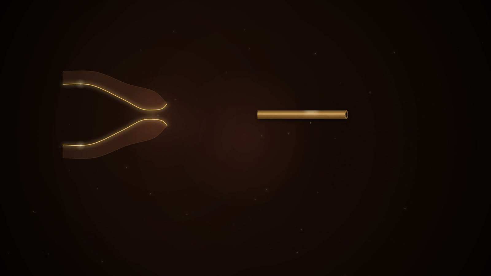
A single gold hollow tube at the right and a glowing gold outline of two curved, tapering walls at the left, on a dark background.

Graduated urethral tubes (Sushruta Samhita)

Period
As described in the Sushruta Samhita (text of disputed date)
In the film
First appears 7:16 in the film, II. The King's Permission

For a narrowed urinary outlet the text prescribes a tube open at both ends, made of iron, wood or shellac. The tube is smeared with clarified butter and introduced gently into the urethra. Every third day it is replaced by a thicker one, so the passage is widened in stages rather than at one sitting. This is graduated dilation, the principle of the later bougie. The same page gives a cutting alternative, an incision on the underside of the penis that spares the perineal raphe; which condition that passage treats is not settled in the sources read.
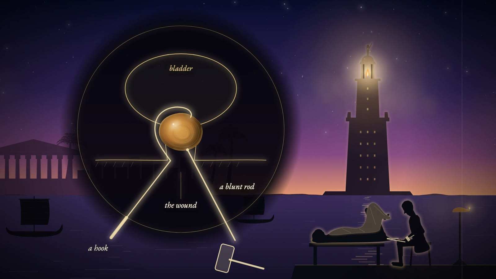
Gold-line bladder with an amber stone gripped by a hook and struck with a blunt rod, beside the Alexandria lighthouse and a figure treating a patient.

Ammonius's hook and splitter

Period
About 200 BC (Riches); described by Celsus
In the film
First appears 10:21 in the film, III. The Oath and the Ligature

When a stone was too large to draw out through the perineal wound, Celsus describes the method credited to Ammonius. A scoop or hook was passed over the stone so that it held the stone steady at the wound, even under a blow. A second instrument of moderate thickness, thin but blunt at its front end, was set against the stone and struck, splitting it. Celsus stresses that the instrument must not touch the bladder and that no fragment of the broken stone may cut into it. The stone was broken instead of the wound being enlarged.

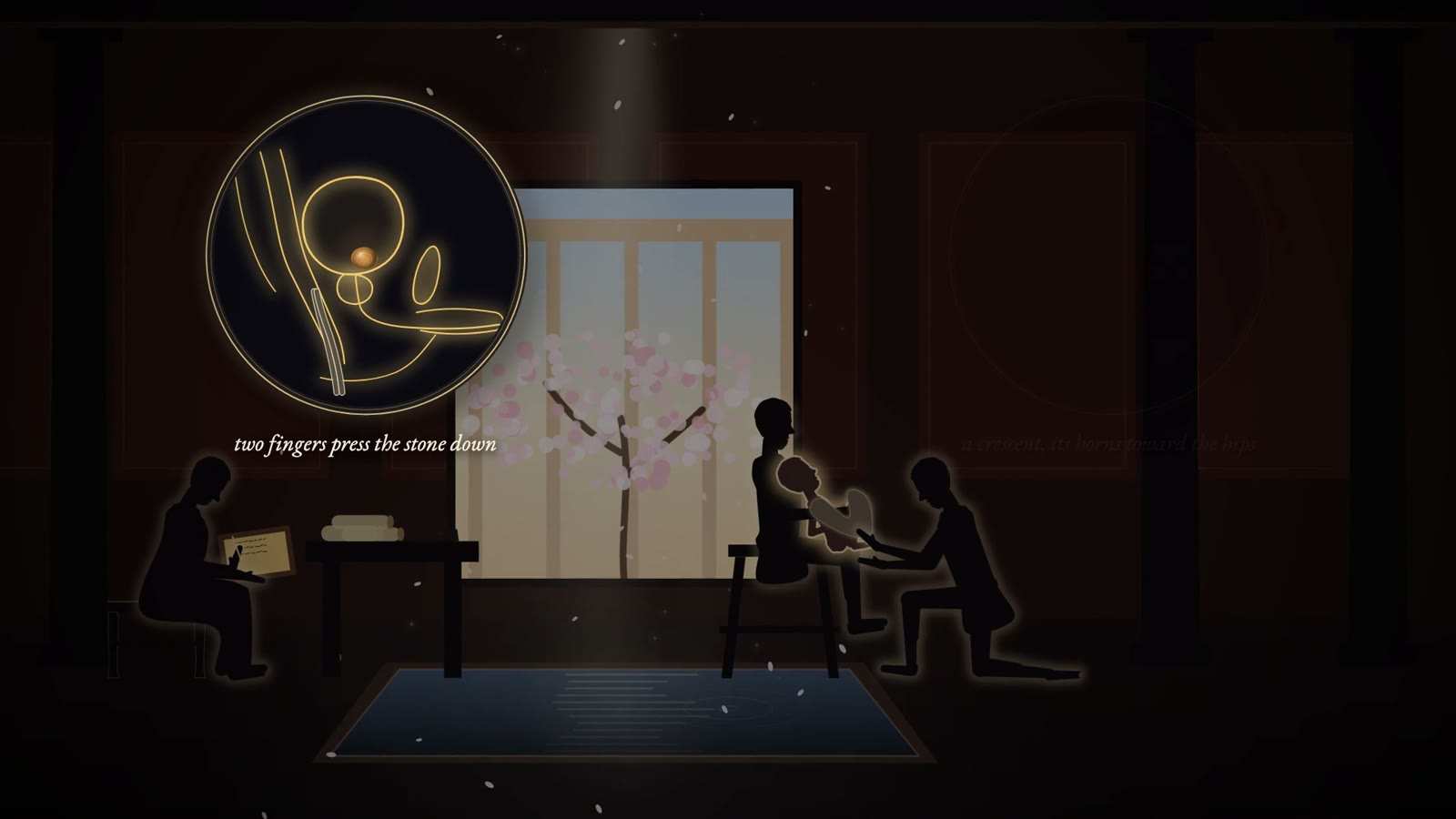
Roman room with a blossoming tree: an assistant holds a boy for a kneeling surgeon, a scribe writes, and an inset reads two fingers press the stone down.

Bronze catheters (Celsus)

Period
Described by Celsus (c. 25 BC to c. AD 50, per Riches); a Pompeii example survives as a 1903 cast
In the film
First appears 11:27 in the film, III. The Oath and the Ligature

For retention of urine Celsus tells the surgeon to keep five bronze tubes ready: three for men, of fifteen, twelve and nine finger-breadths, and two for women, of nine and six. They should be very smooth, neither too wide nor too narrow, and a little curved, more so for men. The tube is passed along the urethra into the bladder to let the urine out. A bronze male catheter found at Pompeii, known from a 1903 cast in the Penn Museum, is 30.5 cm long, close to Celsus's longest size. Celsus does not call the curve an S.

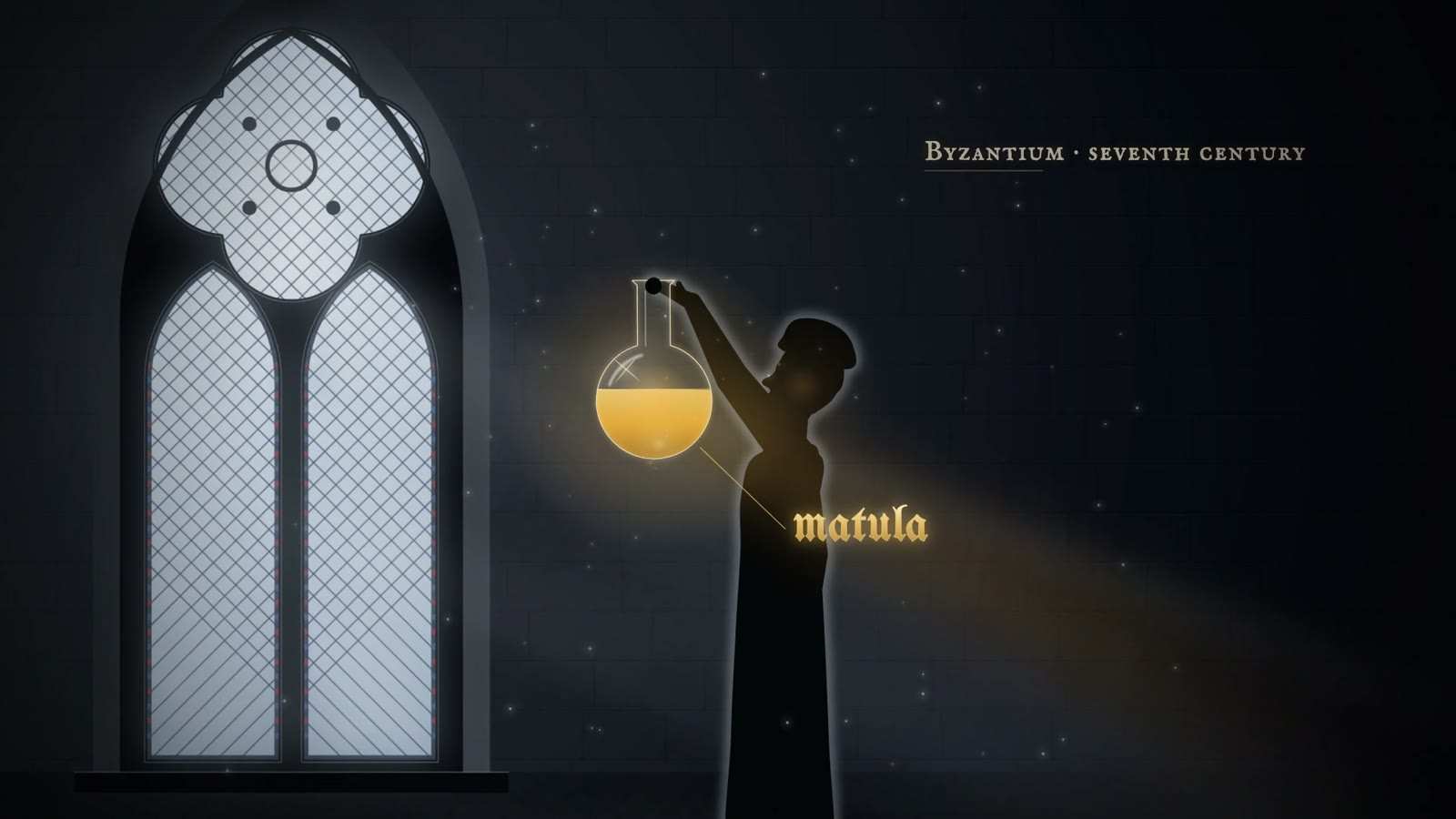
Robed physician in silhouette holds up a round flask of amber urine labelled matula, beside a tall Gothic window, captioned Byzantium, seventh century.

Matula (urine flask)

Period
Byzantine period to the 17th century in learned medicine; asked for by patients into the 19th century
In the film
First appears 14:51 in the film, IV. The Flask

A flask of clear, colourless glass with a round body shaped like a bladder, a rounded base and a narrow mouth. Clear glass let light pass without tinting the specimen, and the rounded base gathered the sediment where it could be inspected. The physician held it at eye level against a window or a candle and judged colour, consistency, quantity, froth and sediment, and sometimes smell. The column of liquid was read in zones, from the ring at the surface down to the sediment. Across medieval Europe the flask became the recognised emblem of the physician.

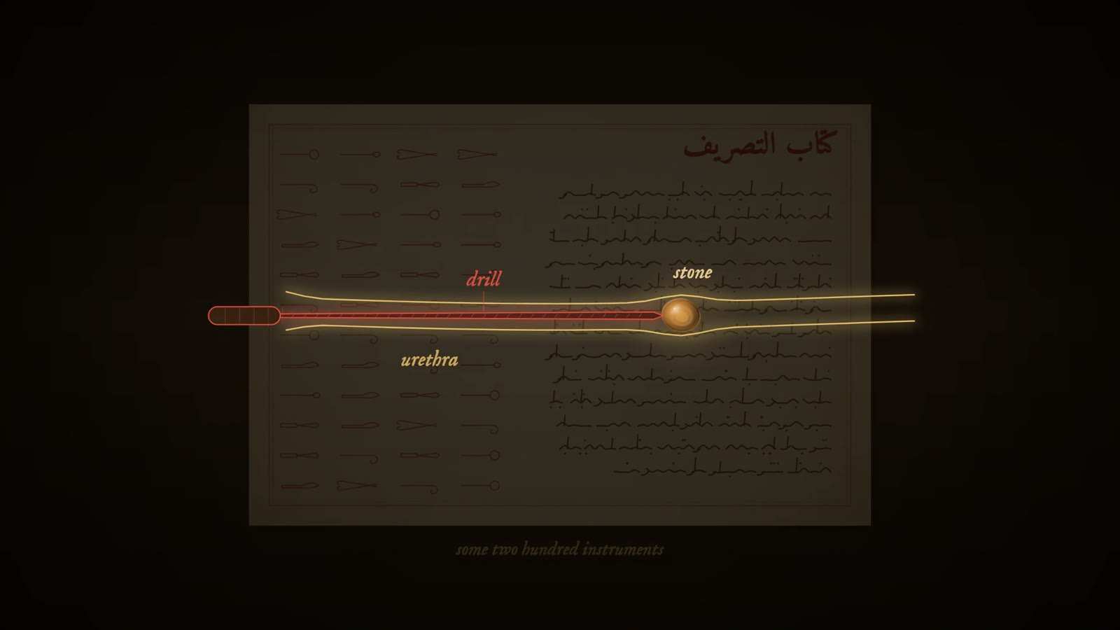
Arabic manuscript page of instrument drawings with a gold-line urethra on it, where a red drill reaches an amber stone, captioned some two hundred instruments.

Bladder syringe (al-Zahrawi) probable

Period
c. 1000, described in al-Tasrif
In the film
First appears 16:35 in the film, IV. The Flask

A syringe for the bladder (zarraqa): a broad cylindrical barrel worked by a piston, ending in a long, narrow cannula with three small holes at the tip. It was made of silver or ivory and was used to put medicine into the bladder; another account says simply that al-Zahrawi used a syringe to irrigate the bladder. The three-holed tip and the materials rest on one modern source.

Arabic manuscript page of instrument drawings with a gold-line urethra on it, where a red drill reaches an amber stone, captioned some two hundred instruments.

Silver catheter (al-Zahrawi) probable

Period
c. 1000, described in al-Tasrif
In the film
First appears 16:35 in the film, IV. The Flask

A straight, narrow, smooth tube of silver with a small funnel at its outer end, about one and a half spans long, roughly 33 cm. It was lubricated before insertion and was used to wash out the bladder. The description comes from one modern account of al-Tasrif; no translation of the text itself was read for the film.

Arabic manuscript page of instrument drawings with a gold-line urethra on it, where a red drill reaches an amber stone, captioned some two hundred instruments.

Urethral stone drill (al-Zahrawi)

Period
c. 1000, described in al-Tasrif
In the film
First appears 16:35 in the film, IV. The Flask

A slender drill with a fine point, for a stone lodged in the urethra. The surgeon turned it gently against the stone until it was pierced through, then squeezed the penis from outside so that the fragments came away with the urine. One modern account describes a steel shaft with a sharp triangular point, set in a wooden handle. It is one of about two hundred instruments drawn in the surgical part of al-Tasrif, alongside forceps for crushing a large stone in the bladder before extraction.

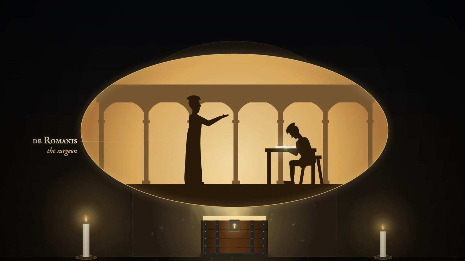
Locked iron-bound chest between two candles, below an oval scene of a robed surgeon reaching toward a seated figure at a desk, labelled the surgeon.

Grooved staff probable

Period
From about 1520 (Marian operation); later adopted by Frère Jacques, Rau and Cheselden
In the film
First appears 27:26 in the film, VI. The Stone-Cutters

A curved metal sound, of silver or steel, with a groove along its convex side and a flat plate for a handle. It was passed along the urethra into the bladder before any cut was made. The surgeon then cut through the perineum down onto the staff and ran the knife along its groove, so that the staff, not touch alone, guided the blade toward the bladder neck. In the Marian operation the conductors, dilators and forceps followed the same track. Frère Jacques first cut without one; his later adoption of a grooved staff, and Rau's, is linked to their better results.

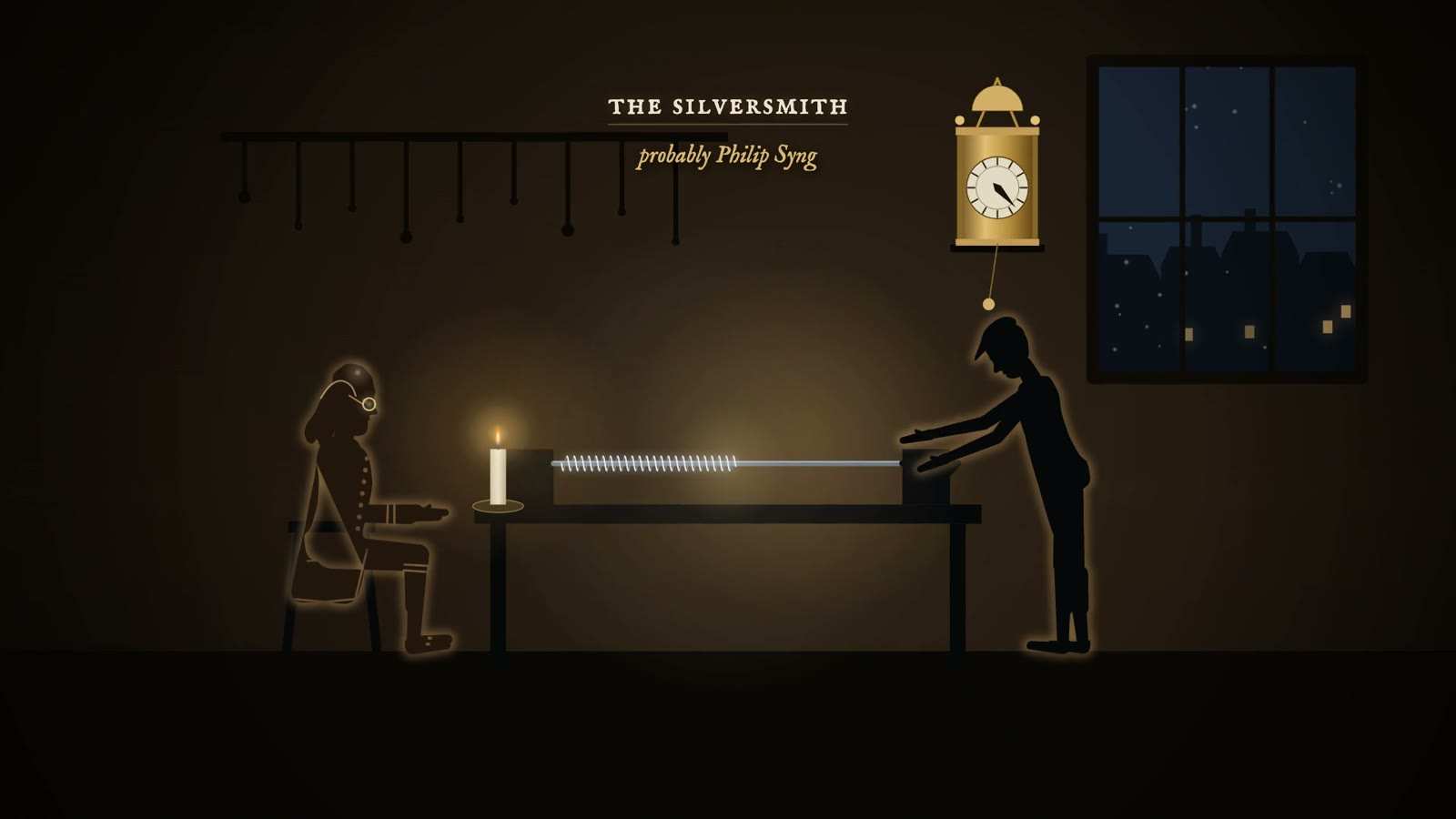
Silversmith at a bench winds a silver wire spiral tube by candlelight while a seated man in spectacles waits, under a wall clock, captioned The Silversmith.

Franklin's flexible silver catheter

Period
1752 (a similar Italian catheter about 1720)
In the film
First appears 33:07 in the film, VI. The Stone-Cutters

Silver wire was wound tightly in a spiral, like a coiled spring, to form a tube, with a short rigid silver pipe at the tip. An inner wire stiffened the back part while it was introduced and was drawn back as the catheter advanced, so the coil could follow the bends of the urethra yet still be pushed forward. Franklin suggested covering it with a thin sheath of gut and greasing it with tallow, and noted that it could be screwed in and out by turning. Roncalli-Parolino's earlier version was a silver spiral covered with fine silk.

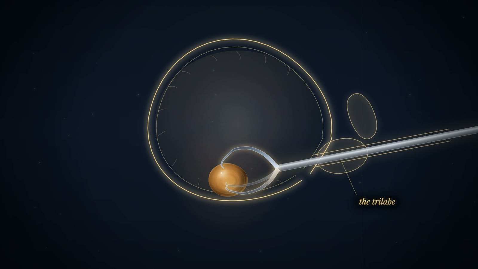
Gold-line bladder with an amber stone held by three curved claws on a straight steel tube entering from the right, labelled the trilabe.

Civiale's trilabe

Period
From 1824
In the film
First appears 33:47 in the film, VII. Breaking the Stone

A straight steel tube, about the length of a forearm, was passed along the urethra into the bladder. Inside the bladder three curved spring branches were pushed out of its end and opened like a claw, then closed on the stone to hold it. A central steel rod with a drill tip ran down the tube and bored into the stone held in the claws, breaking it into pieces that could pass in the urine or be broken further at later sittings. Instrument sets of this kind were made by the Paris maker Joseph Charrière. How the drill was turned is not verified in the kit.

Sources
  1. Riches 1967, The history of lithotomy and lithotrity (Arnott Demonstration, RCS) Secondary
  2. EAU History Office, Exhibit of the month: the Civiale set Museum or society
Gold-line bladder with an amber stone held by three curved claws on a straight steel tube entering from the right, labelled the trilabe.

Heurteloup's percuteur probable

Period
Presented 1832
In the film
First appears 33:47 in the film, VII. Breaking the Stone

A curved-beaked steel lithotrite with two blades. It was passed into the bladder and the stone was caught between the blades. With the instrument held fixed, the operator struck its outer end with a small hammer, so that the blow cracked the stone instead of a drill boring into it. Heurteloup took the method to England, where he was the first to perform lithotrity.

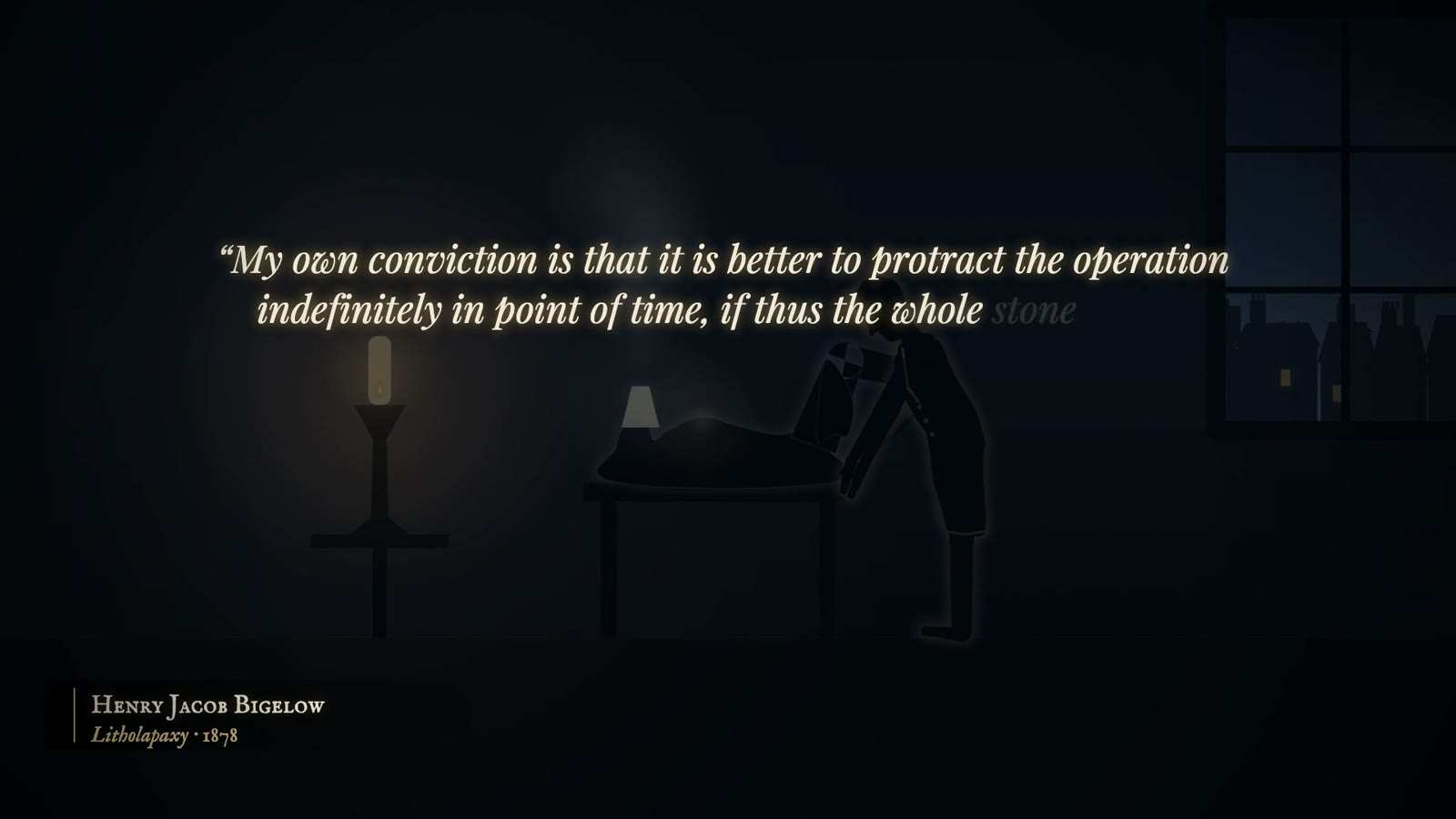
Dim room with a surgeon bending over a patient beside a candle, under a quotation beginning My own conviction is that it is better to protract the operation.

Bigelow's evacuator

Period
1878
In the film
First appears 37:14 in the film, VII. Breaking the Stone

A rubber bulb holding about ten ounces of water was joined to a wide metal evacuating catheter by a length of rubber tube, six inches to two feet long. After the lithotrite had crushed the stone, the operator squeezed and released the bulb so that water washed in and out of the bladder, carrying the fragments with it. The fragments sank to the bottom of the bulb and collected in a glass chamber beneath it. The whole stone could therefore be removed at one sitting instead of being left to pass over weeks.

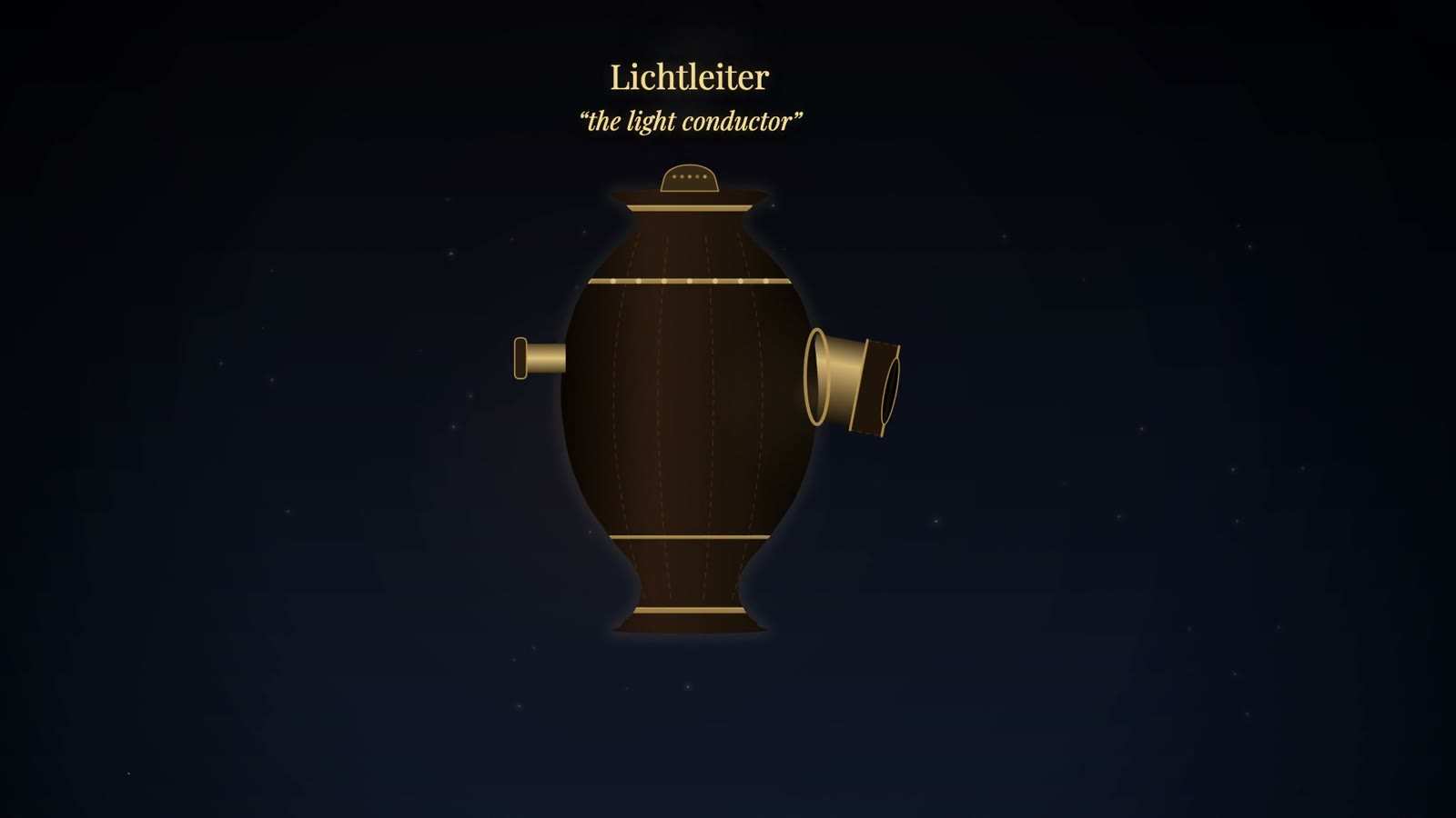
Vase-shaped brass-banded lamp with a short tube on each side, titled Lichtleiter, the light conductor, on a dark starry ground.

Lichtleiter (light conductor)

Period
First shown 1804; described 1806 and 1807
In the film
First appears 39:07 in the film, VIII. Light

A vase-shaped housing about 35 cm tall held a wax candle on springs, so that the flame kept its position as it burned down, in front of a concave mirror. A round port on the front was divided by a partition: one half sent light down an attached tube into the cavity, the other carried the image back to an eyepiece set off-centre on the back wall, so that the flame's glare did not swamp the view. Interchangeable tubes and four-bladed, screw-spread specula attached through a metal sleeve and a leather cuff, for large cavities, small openings, or an oblique view through a mirror at 90 degrees. The covering is disputed: leather over lead, paper and leather over sheet metal, or sharkskin over silver.

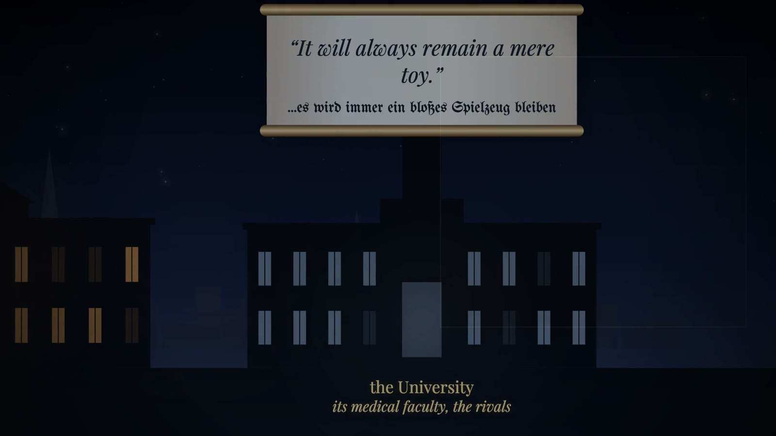
Scroll above a night-time university building reading It will always remain a mere toy, in English and German; caption the University, its medical faculty.

Ilg's Viennese model of the Lichtleiter probable

Period
Built late 1806 to January 1807; no original survives
In the film
First appears 39:43 in the film, VIII. Light

Johann Georg Ilg, prosector of the Josephinum, rebuilt Bozzini's instrument within two months. He used two candles, a single channel and a perforated mirror, which the EAU describes as the first use of 'Gregory's principle' in an instrument. His specula opened all four blades with one disc, where Bozzini's had to be set one by one. At an Academy session on 17 January 1807 it was judged superior for ease of use and brightness. Karl Storz built a replica in 2004 from Ilg's report of 1807.

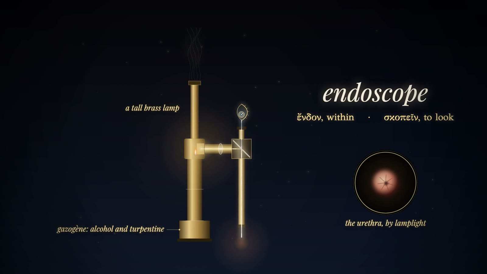
Tall brass lamp, a lens and a mirror block feeding a vertical tube with an eye at the top, labelled endoscope, with an inset of the urethra by lamplight.

Desormeaux's endoscope

Period
Shown 1853; described 1865 (English translation 1867)
In the film
First appears 40:51 in the film, VIII. Light

A lamp burning gazogene, a mixture of alcohol and turpentine, stood beside the instrument under a chimney. A concave mirror behind the flame and a plano-convex lens in front concentrated the light onto a mirror set obliquely at the top of a straight metal tube. The mirror was pierced at its centre, and the examiner looked through the hole down the tube, so light and line of sight shared one axis. Desormeaux wanted a flame that was intense but small, and judged candles, oil and petroleum useless. He used it to inspect the urethra and bladder, to cut strictures and to cauterise lesions under vision. The maker was Charrière of Paris.

Tall brass lamp, a lens and a mirror block feeding a vertical tube with an eye at the top, labelled endoscope, with an inset of the urethra by lamplight.

Ségalas's urethro-cystic speculum

Period
Presented 1826; clinical cases reported 1826 to 1828
In the film
First appears 40:51 in the film, VIII. Light

Two silver tubes, two metal mirrors and two small candles held in the examiner's left hand, with an elastic probe. The urethral tube was polished inside and widened into a conical mirror, and a hardened rubber obturator eased insertion. The design was concentric: a central viewing tube, its eye funnel blackened inside, with the light carried around it. There was no fixed lamp housing, and the open candles risked burns. Only three clinical cases were reported. According to Ségalas, Fresnel told him after the presentation to put the light at the side and reflect it down the tube.

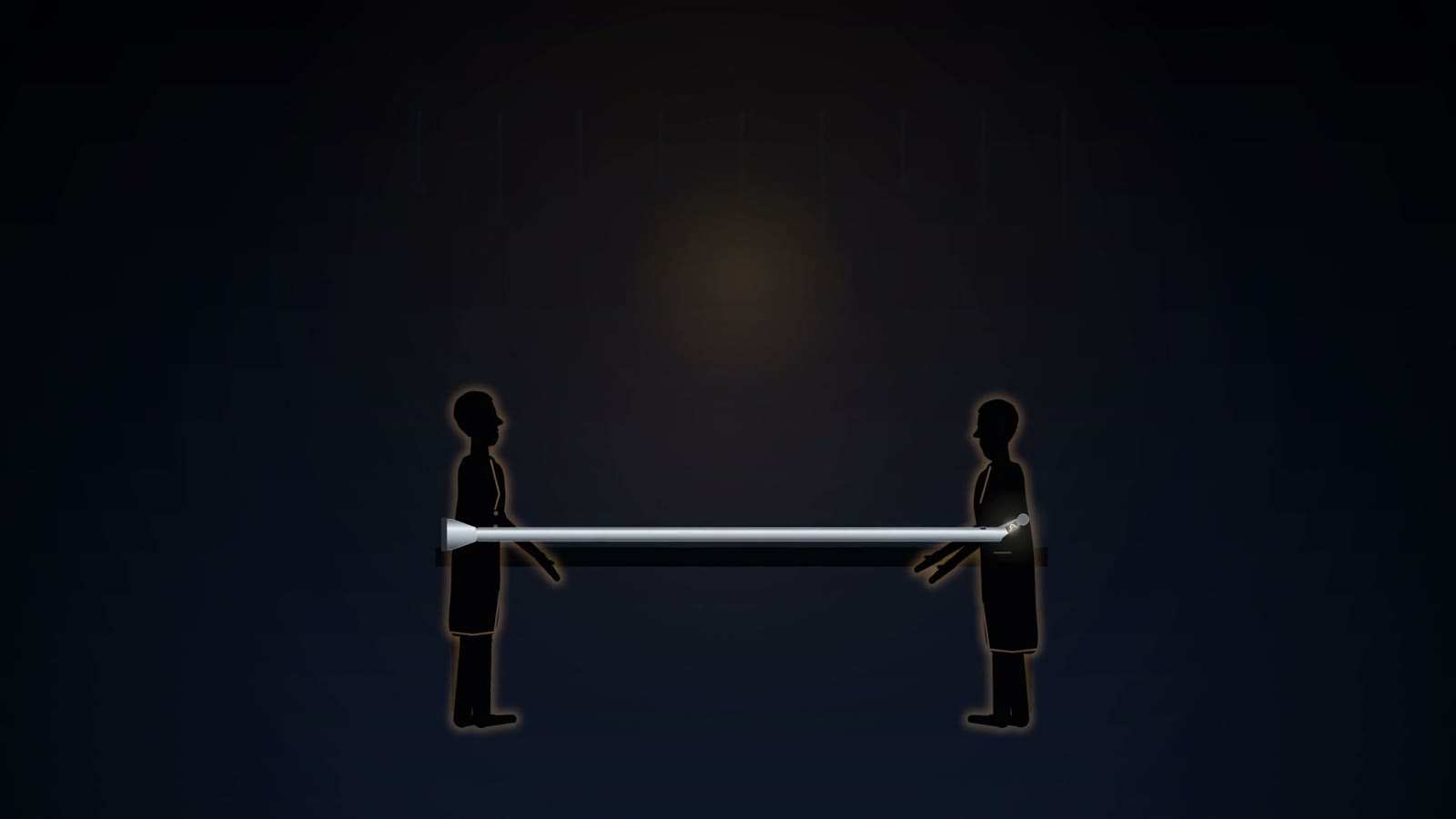
Two silhouetted men face each other holding a long, slim metal shaft between them, flared at the left end and with a small jointed tip at the right.

Nitze-Leiter cystoscope (1879)

Period
Shown on a living patient 9 May 1879; superseded by lamp cystoscopes from 1886-1887
In the film
First appears 42:02 in the film, VIII. Light

A slim, catheter-shaped metal shaft carried its own light at the tip: a platinum wire, made white-hot by battery current, behind a small flat window. The filament became very hot and burnt out easily, so iced water ran continuously through channels in the shaft to carry the heat away. Inside the shaft a telescopic system of lenses gave a wide field, and because the light travelled with the tip the bladder was lit however the instrument was moved. Tubes for the water and leads to a galvanic battery ran from the eyepiece end. Many early instruments gave a mirror-reversed image until prisms corrected it.

Two silhouetted men face each other holding a long, slim metal shaft between them, flared at the left end and with a small jointed tip at the right.

Incandescent-lamp cystoscope

Period
Late 1886 to 1887 (Leiter with Dittel in Vienna; Nitze with Hartwig in Berlin)
In the film
First appears 42:40 in the film, VIII. Light

A small carbon-filament lamp replaced the platinum wire. In Leiter's version, as Newell described it in 1887, the shaft was two thin metal tubes separated by insulation; the lamp sat in a screw-on lamp-house with a thick crystal window in its side, and a switch at the eyepiece end turned it on only after insertion. The bladder was filled with six to eight ounces of fluid, which carried off the heat, so water cooling was no longer needed. The lamps ran at about three to four volts from a plunge battery and lasted about thirty hours. Fenwick preferred Leiter's replaceable cartridge lamp to Nitze's screw-on unit.

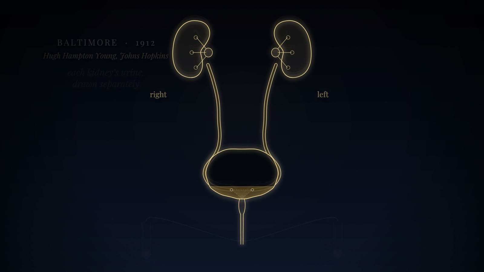
Gold-line kidneys, ureters and bladder with both ureteric openings marked, labelled right and left, with the caption Baltimore, 1912.

Albarrán's deflecting lever probable

Period
c. 1897
In the film
First appears 43:24 in the film, VIII. Light

A small hinged metal flap, the levier or onglet d'Albarrán, sat at the tip of the cystoscope over the channel carrying a thin ureteral catheter. Raising it, by a wheel at the eyepiece end, bent the catheter towards the ureteric orifice in the field of view, so the operator could steer the catheter into the ureter instead of angling the whole instrument. Albarrán adapted the device to the cystoscopes of Casper and Nitze. The dossier's account is functional; the film draws the lever deflecting downward with the scope rotated through 180 degrees, following the commissioning urologist's description, not a period drawing.

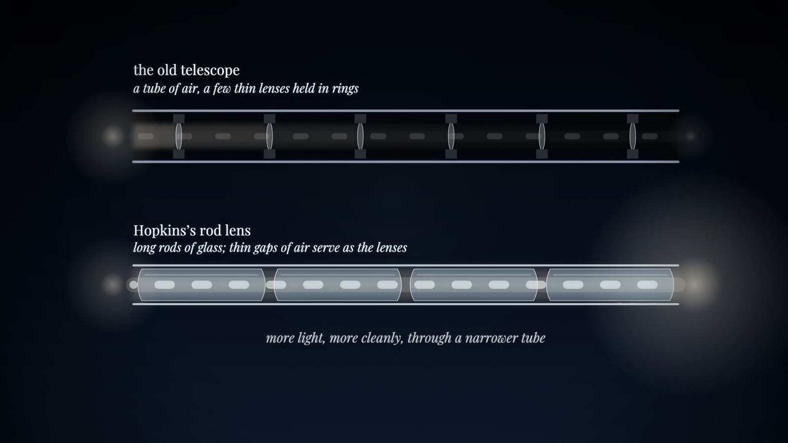
Two tube diagrams: the old telescope with a few thin lenses, and the rod lens of long glass rods, captioned more light, more cleanly, through a narrower tube.

Hopkins-Kapany fibrescope

Period
Published January 1954
In the film
First appears 46:57 in the film, VIII. Light

One continuous glass fibre was wound in a figure of eight round two drums, then bound, cut and polished. Because every fibre kept its place relative to its neighbours along the whole length, the bundle was coherent: an image focused on one end reappeared at the other as a pattern of points, even when the bundle was bent. Bundles of more than 10,000 fibres carried images along about 75 cm. The first fibres were unclad and leaked light where they touched; glass cladding, from van Heel's idea and Curtiss's glass-clad fibres, solved that problem.

Two tube diagrams: the old telescope with a few thin lenses, and the rod lens of long glass rods, captioned more light, more cleanly, through a narrower tube.

Hopkins rod-lens telescope with Storz cold light

Period
Patent application 1959; rod-lens cystoscope with cold light from 1967
In the film
First appears 46:57 in the film, VIII. Light

The older endoscope telescope was a thin tube that was mostly air, with small thin lenses held in metal rings that blocked part of each lens. Hopkins reversed the arrangement: long glass rods with convex ends filled most of the tube, and the short spaces of air between them acted as the lenses. The rods fill the bore and centre themselves, so the usable aperture is larger, and fewer glass-air surfaces lose less light. Storz paired the telescope with an external cold-light source whose light reached the tip through a bundle of glass fibres. Secondary accounts put the gain in light at up to about eighty times.

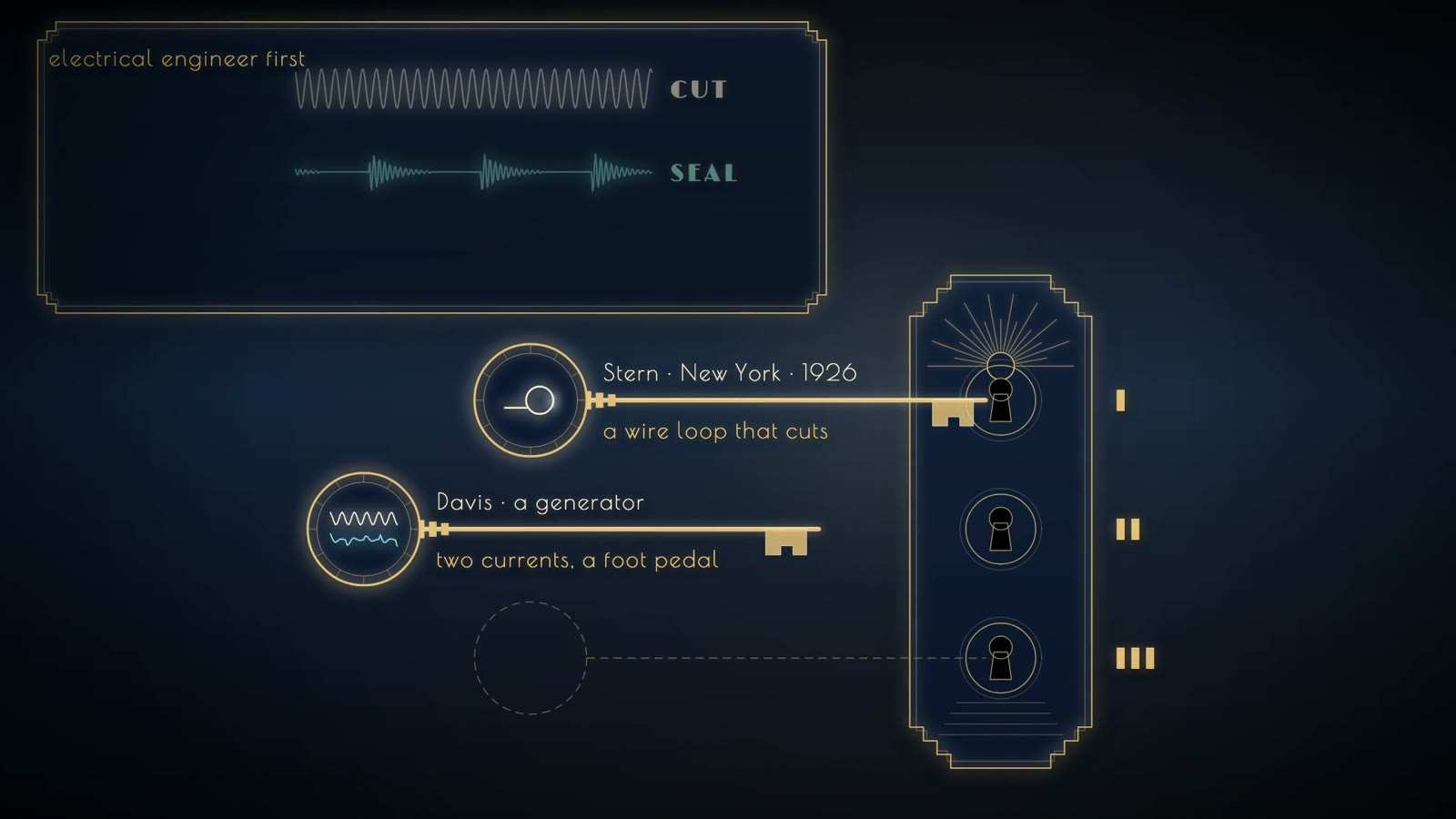
Lock with three keyholes; keys labelled Stern, New York, 1926, a wire loop that cuts, and Davis, a generator with two currents and a foot pedal; third empty.

Bottini's galvanocautery incisor

Period
1874
In the film
First appears 51:07 in the film, IX. The Gland That Stands Before

A transurethral instrument for an obstructing prostate, devised by Enrico Bottini in Pavia. It was passed along the urethra with no means of seeing, and carried a blade heated by electric current, with which the surgeon burned an incision through the prostate and bladder neck. The operator worked by feel and position alone. Later histories treat it as a distant ancestor of transurethral resection. The kit confirms the electrically heated blade, the place and the year; the instrument's exact shape and power source should be checked against a museum example before it is described further.

Lock with three keyholes; keys labelled Stern, New York, 1926, a wire loop that cuts, and Davis, a generator with two currents and a foot pedal; third empty.

Davis-Bovie electrosurgical generator

Period
about 1931
In the film
First appears 51:07 in the film, IX. The Gland That Stands Before

A generator built by Theodore Davis with G. H. Liebel that supplied two kinds of current: a high-frequency current that cut, and a lower-frequency current that coagulated bleeding vessels. The surgeon switched between them electromagnetically with foot switches, keeping both hands on the resectoscope. Davis showed films of resections done with it at the 1931 meeting of the American Urological Association in Memphis, and reported 966 resections with 7 deaths. The appearance of the cabinet is not documented in the kit.

Lock with three keyholes; keys labelled Stern, New York, 1926, a wire loop that cuts, and Davis, a generator with two currents and a foot pedal; third empty.

Stern-McCarthy resectoscope

Period
1931 onward
In the film
First appears 51:07 in the film, IX. The Gland That Stands Before

Joseph McCarthy's instrument of 1931 brought together Stern's cutting loop, Davis's two currents and McCarthy's own panendoscope with a Foroblique (forward-oblique) lens, made with the Wappler firm of American Cystoscope Makers. All of it sat in a Bakelite sheath that insulated the patient from the current. A rack-and-pinion mechanism drew the loop back toward the operator, as in resection today; one surviving example has an ebony handle. It became the standard resectoscope internationally.

Lock with three keyholes; keys labelled Stern, New York, 1926, a wire loop that cuts, and Davis, a generator with two currents and a foot pedal; third empty.

Stern's resectoscope

Period
January 1926
In the film
First appears 51:07 in the film, IX. The Gland That Stands Before

Maximilian Stern's instrument, presented to the New York Academy of Medicine in January 1926, carried a tungsten wire loop through which an undamped high-frequency current was passed. Drawn through the obstructing tissue, the electrified loop cut away strips of prostate. It cut well but coagulated poorly, so bleeding remained the main difficulty. That weakness was met by Davis's two-current generator and by McCarthy's redesign of 1931.

Lock with three keyholes; keys labelled Stern, New York, 1926, a wire loop that cuts, and Davis, a generator with two currents and a foot pedal; third empty.

Young's prostatic punch

Period
1909 (cautery added 1911)
In the film
First appears 51:07 in the film, IX. The Gland That Stands Before

A modified urethroscope with a lengthened beak and a window in its side. Obstructing tissue that bulged into the window was cut off by a tubular knife pushed down the shaft. Hugh Hampton Young introduced it on 1 April 1909, after a patient's death, and used it under cocaine local anaesthesia; cautery to control bleeding was added in 1911. Later punches by Caulk (1920, with an insulated sheath) and Kenneth Walker (1925, with a Bakelite sheath, telescope and continuous irrigation) refined the idea before the loop resectoscope replaced it.

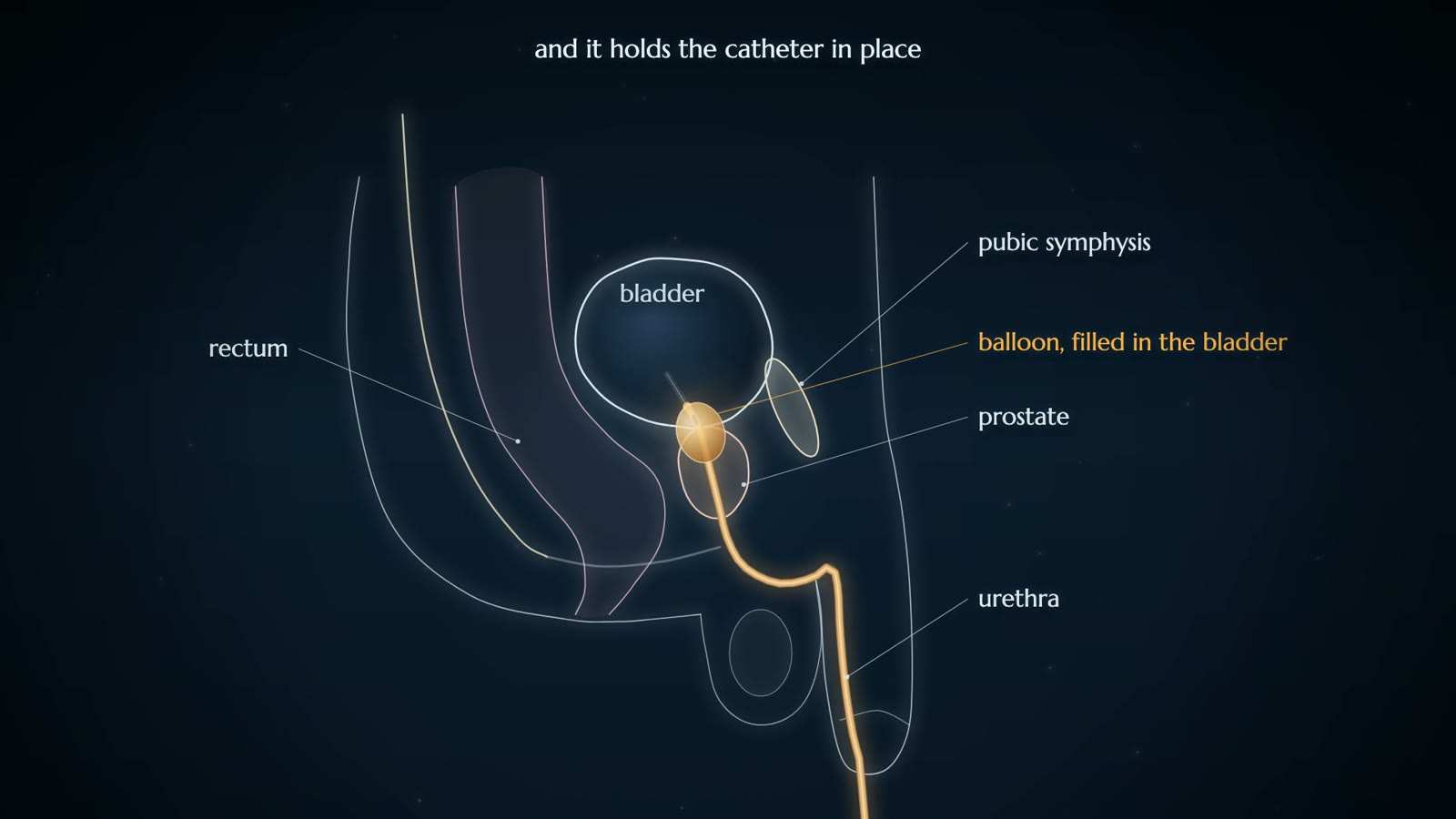
Side-view diagram of the male pelvis with a catheter running up the urethra and a filled balloon in the bladder, labelled rectum, prostate and pubic symphysis.

Foley balloon catheter

Period
first described 1929; published 1937
In the film
First appears 57:49 in the film, X. Without the Knife

A one-piece latex rubber catheter with an inflatable bag, or balloon, near its tip. Once the catheter was in the bladder the balloon was filled, so the catheter held its place and drained continuously without outside fixation. Foley first described it in 1929 to control bleeding after prostatic resection, demonstrated it in 1935 and published it in 1937 as a haemostatic bag catheter. C. R. Bard sold the devices as Foley catheters from 1935, although the patent went to Paul Raiche of the Davol Rubber Company.

Sources
  1. Wikipedia: Frederic Foley Secondary
  2. Wikipedia: Foley catheter Secondary
  3. F. E. B. Foley, A hemostatic bag catheter: one piece latex rubber structure for control of bleeding and constant drainage following prostatic resection, Journal of Urology 1937; 38:134-139 (title and citation as given on Wikipedia) Primary
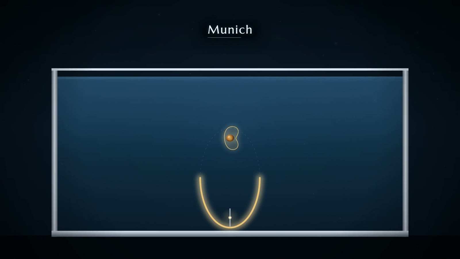
Water tank captioned Munich, with a gold curved reflector at its base and a kidney holding an amber stone above it, joined by a faint dashed ellipse.

Dornier lithotripter (HM1 to HM3)

Period
HM1 prototype 1979; first patient 7 February 1980; HM3 from 1983
In the film
First appears 1:00:03 in the film, X. Without the Knife

The patient lay in a tub of water, which carried the shock wave into the body without an air gap. Below the flank, an underwater spark discharge at the first focus of a half-ellipsoid reflector produced a shock wave, and the reflector gathered it onto the second focus, where the stone had been placed using two X-ray systems set in two planes. Repeated shocks broke the stone into fragments small enough to pass. Early patients had spinal anaesthesia, but peridural anaesthesia soon proved enough. The HM3 was the first commercial machine.

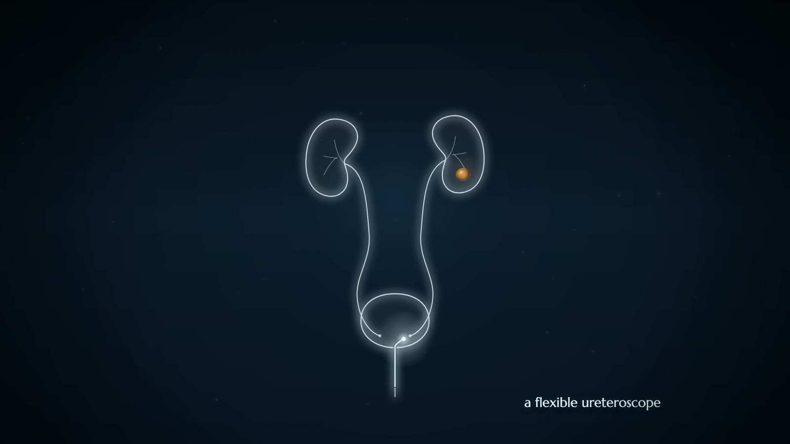
Outline of two kidneys, ureters and bladder, an amber stone in one kidney and a thin scope entering the bladder from below, captioned a flexible ureteroscope.

Holmium:YAG laser for lithotripsy probable

Period
laser lithotripsy from the 1980s; holmium the standard from the mid-1990s
In the film
First appears 1:00:55 in the film, X. Without the Knife

Laser light is delivered in pulses onto a stone through a fibre passed along an endoscope, breaking the stone so that it can be removed or passed. Laser lithotripsy was developed at Massachusetts General Hospital in the 1980s, first with pulsed dye lasers at 504 nm. Holmium:YAG lasers, working in the infrared at about 2,100 nm, were studied in the 1990s and became the standard from the mid-1990s; thulium fibre lasers spread in the present century. The first clinical year of holmium use is not verified in the kit.

Sources
  1. Wikipedia article on laser lithotripsy, as cited in research/03 §16 (URL not recorded in the kit) Secondary
Outline of two kidneys, ureters and bladder, an amber stone in one kidney and a thin scope entering the bladder from below, captioned a flexible ureteroscope.

Purpose-built rigid ureteroscope probable

Period
1980
In the film
First appears 1:00:55 in the film, X. Without the Knife

A telescope made to pass up the ureter to a stone. Earlier looks into the upper tract had used instruments built for other purposes. In 1980 Pérez-Castro, working with Karl Storz, produced a rigid ureteroscope 12 French in calibre and 50 cm long, with a separate optic and a working channel for instruments, and used it to treat a kidney stone the following year. Semi-rigid ureteroscopes followed in 1989; flexible ureteroscopes gained active deflection and working channels during the 1980s, and later carried a camera chip at the tip.

Sources
  1. L. A. Whitehurst, B. K. Somani, Urolithiasis 2018 (history of ureteroscopy) Secondary
  2. Bagley, History of ureteroscopy (Springer chapter) Secondary
Hospital building in silhouette at night with lit windows and golden sparks gathering over its centre, captioned Henry Ford Hospital, Detroit.

da Vinci surgical system

Period
cleared 11 July 2000; single-port version cleared 2018
In the film
First appears 1:01:15 in the film, X. Without the Knife

As the film describes robotic surgery, the surgeon sits at a console away from the table while mechanical arms hold and move the instruments inside the patient. The kit's verified details for this system are regulatory. Intuitive Surgical was founded in 1995; the FDA cleared the da Vinci for general laparoscopic surgery on 11 July 2000, with a further clearance on 30 May 2001 listed under gastroenterology and urology. The single-port da Vinci SP was cleared in 2018 only for urological procedures suited to a single port. The kit's descriptions of carts and consoles are generic.