26 operations, in order of appearance

Operations

History, not technique: a plain account of how each operation was done in its period, and the moment it first appears in the film.

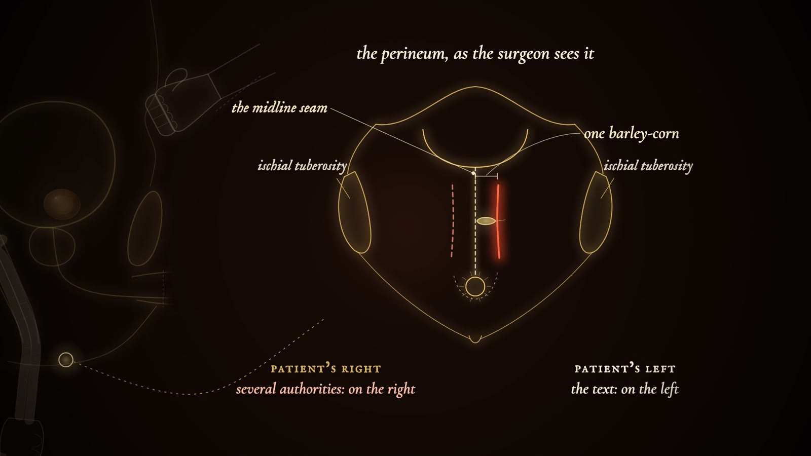
Gold-line diagram titled the perineum, as the surgeon sees it, with midline seam, ischial tuberosities, one barley-corn and a red incision on the left.

Perineal lithotomy (Sushruta Samhita)

Period
Sushruta Samhita, Chikitsita Sthana ch. VII (text of disputed date)
In the film
First appears 5:58 in the film, II. The King's Permission

Medicines were tried first. If they failed, and with the king's permission, the surgeon passed the second and third fingers of the left hand into the rectum and brought the stone forward between rectum and penis, while a closed fist pressed the left side of the umbilical region to hold it within reach. An incision was made on the left of the perineal raphe, a barley-corn's breadth from it, wide enough for the stone to pass, and the stone was taken out. Several authorities cut on the right. Afterwards the patient sat in a cauldron of warm water, took a treacle drink, and the wound was dressed with honey and clarified butter.
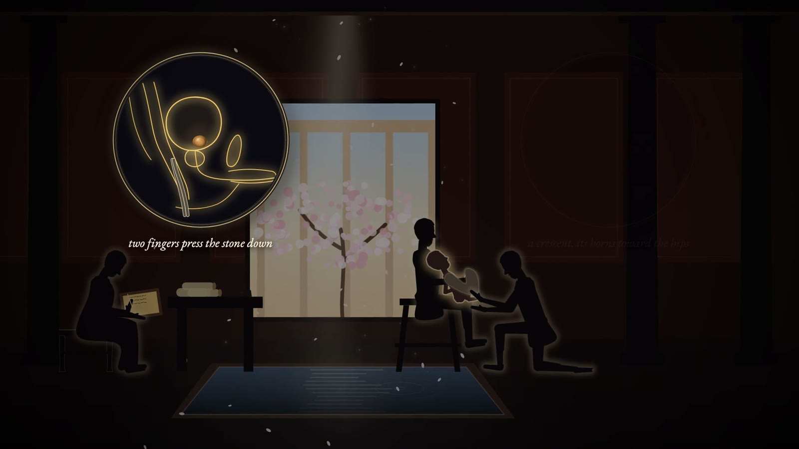
Roman room with a blossoming tree: an assistant holds a boy for a kneeling surgeon, a scribe writes, and an inset reads two fingers press the stone down.

Celsian lithotomy (the lesser apparatus)

Period
Described by Celsus (c. 25 BC to c. AD 50, per Riches); standard in Europe until the Marian operation of the 1520s and 1530s
In the film
First appears 10:53 in the film, III. The Oath and the Ligature

Celsus reserves the operation for spring and for boys aged nine to fourteen whose disease cannot otherwise be relieved. A strong assistant on a high stool holds the child on his knees, back to the assistant's chest, while the child draws his knees up with his hands. The surgeon, nails pared, passes the index and middle fingers of the left hand into the anus, finds the stone at the bladder neck and presses it toward the perineum, the right hand pressing from above. A crescent-shaped incision is made over the stone near the anus, its horns toward the hips, and the stone is removed. Few instruments were needed, hence the later name apparatus minor.

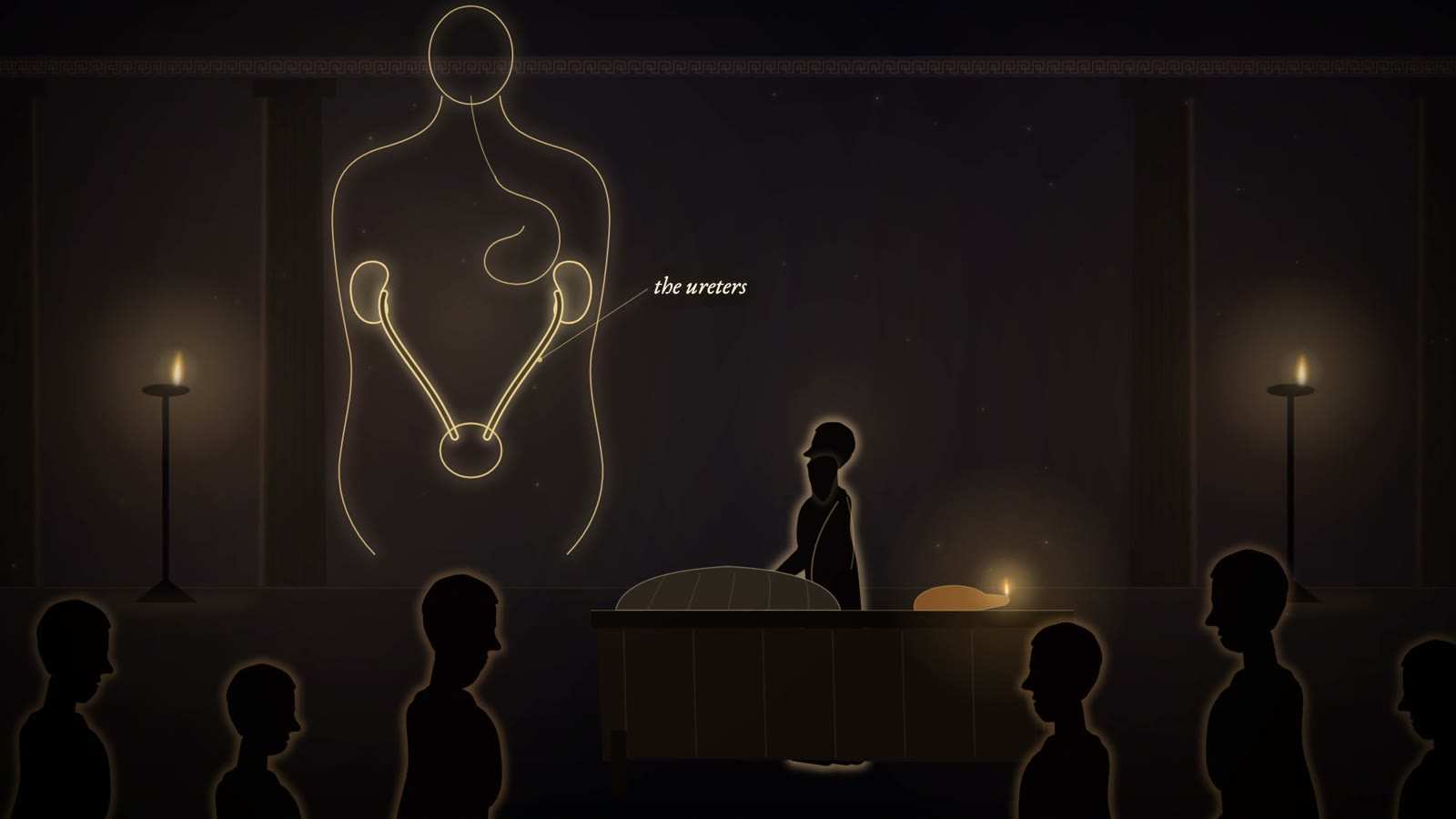
Galen at a demonstration table before spectators in togas, beside a gold-line torso showing kidneys, ureters and bladder, with the ureters labelled.

Galen's ureteric ligation experiment

Period
2nd century AD; On the Natural Faculties I.13
In the film
First appears 12:42 in the film, III. The Oath and the Ligature

Galen opened the peritoneum in front of both ureters of a living male animal, tied them, bandaged the animal and let it go; it stopped passing urine. On unbandaging, the bladder was empty and the ureters were distended. With the ligatures removed, the bladder filled. With the penis tied, squeezing the full bladder sent nothing back up the ureters. With one ureter tied and the other free, only the free side filled the bladder. Cutting the distended ureter made urine spurt like blood at venesection. After both were cut, the bladder stayed empty and urine collected in the abdomen. The kidneys, not vapour, supply the bladder.

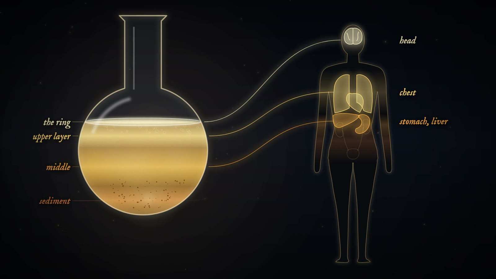
Round flask of layered urine labelled the ring, upper layer, middle and sediment, linked by lines to a human outline lit at head, chest, and stomach and liver.

Uroscopy (reading the urine in the flask)

Period
Seventh to seventeenth century as the central test of learned medicine; popular until the 19th century
In the film
First appears 14:51 in the film, IV. The Flask

The physician held the matula to the light and judged colour on a scale of about twenty shades, from clear to black, together with consistency, quantity, froth, sediment and smell, and sometimes taste. The column of liquid was read in layers mapped onto the body: the ring at the surface for the head and senses, the upper layer for the chest, the middle for the stomach, liver and spleen, and the sediment for the kidneys, bladder, uterus and genitals. Within humoral medicine the reading guided both diagnosis and treatment. Often the urine came by messenger and the patient was never seen.

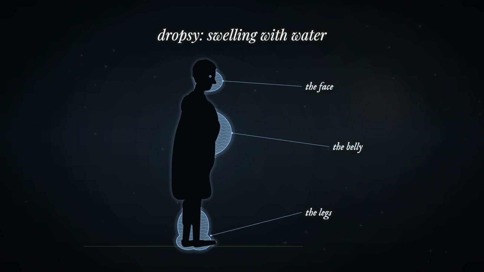
Silhouette of a standing figure in profile with blue water-filled swellings at the face, belly and legs, titled dropsy: swelling with water.

Heat test for coagulable urine (Bright)

Period
1820s (Bright, Reports of Medical Cases, 1827), after earlier observations by Dekkers and Cotugno
In the film
First appears 20:41 in the film, V. What the Water Knew

A little urine was put in a spoon and held over a candle flame. In Bright's words, before it rose quite to the boiling point it became clouded: the urine was coagulable. Earlier, Dekkers and Cotugno had seen urine coagulate with heat or acetic acid; Cotugno described a dense white body like egg albumen in the urine of a soldier with dropsy. Bright joined the bedside test to the post-mortem table, finding diseased kidneys in patients who had died with dropsy and coagulable urine.

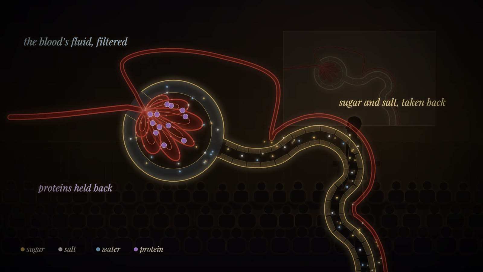
Diagram of a kidney tuft in its cup and winding tubule, labelled the blood's fluid, filtered; proteins held back; and sugar and salt, taken back.

Glomerular micropuncture (Wearn and Richards)

Period
First reported December 1921; full paper 1924
In the film
First appears 24:53 in the film, V. What the Water Knew

A frog was anaesthetised and its kidney exposed under a microscope and an intense light. Using a micromanipulator, a technique the team had seen Robert Chambers demonstrate in December 1920, a fine glass micropipette was pushed into the capsule of a single glomerulus and fluid was drawn from the space inside. Bladder urine was collected at the same time. Silver nitrate showed chloride and Benedict's solution showed sugar in the capsular fluid but not in the bladder urine, and no protein was detected in eleven glomerular samples.

Sources
  1. Schmidt 1971, NAS Biographical Memoir of Alfred Newton Richards Secondary
  2. Wearn JT, Richards AN. Observations on the composition of glomerular urine, with particular reference to the problem of reabsorption in the renal tubules. Am J Physiol 1924;71(1):209-227. doi:10.1152/ajplegacy.1924.71.1.209 Primary
  3. Dirks, Clapp and Berliner 1964 (J Clin Invest) Primary
  4. Vallon 2008, Micropuncturing the nephron Secondary
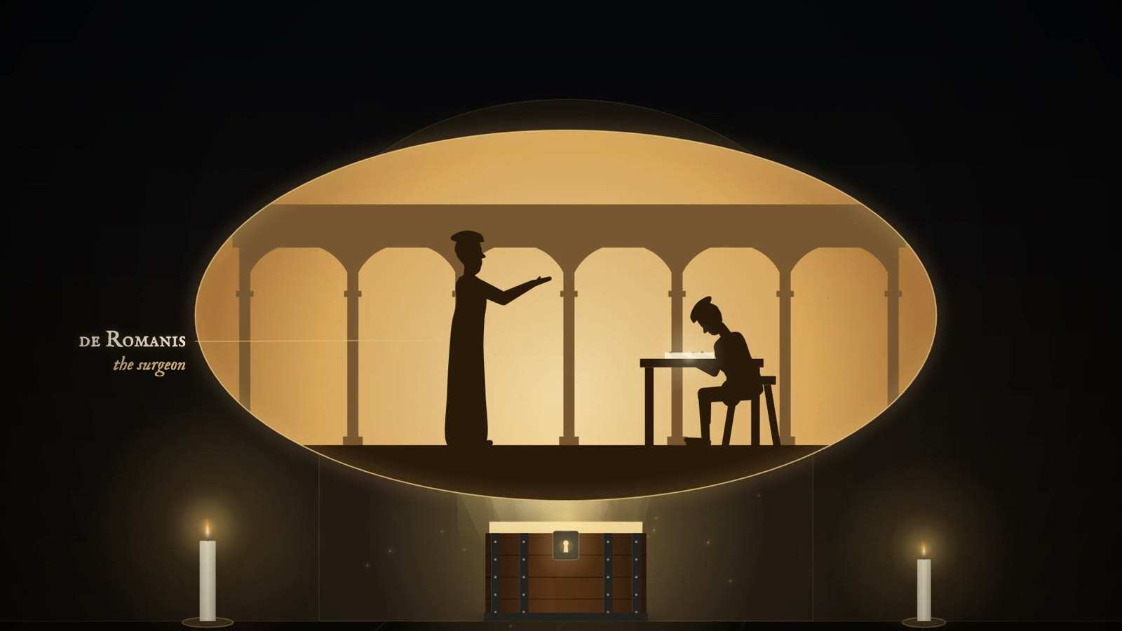
Locked iron-bound chest between two candles, below an oval scene of a robed surgeon reaching toward a seated figure at a desk, labelled the surgeon.

Marian operation (the greater apparatus) probable

Period
Devised about 1520 by de Romanis; published by Mariano Santo (1522 per Riches; the Libellus aureus, 1535)
In the film
First appears 27:26 in the film, VI. The Stone-Cutters

The reasoning was that stones pass easily through the short, dilatable female urethra, so a male urethra made shorter and wider might let them through too. A grooved staff was passed down the urethra into the bladder and the surgeon cut onto it in the perineum. Conductors, dilators and long-handled toothed forceps were then passed along the staff to open the way and draw out the stone. The crowded tray gave the name apparatus major, against the Celsian apparatus minor, in which the surgeon cut onto a stone pressed down by fingers in the rectum. The Colot family were among the lithotomists who adopted it.

Gold-line side view of a child's pelvis with an amber stone in the bladder, labelled bladder, peritoneum, symphysis and rectum, captioned the Hippocratic rule.

Suprapubic lithotomy (the high operation) probable

Period
Franco, 1550s or 1561; revived in London by John Douglas (1719) and Cheselden (1723)
In the film
First appears 28:07 in the film, VI. The Stone-Cutters

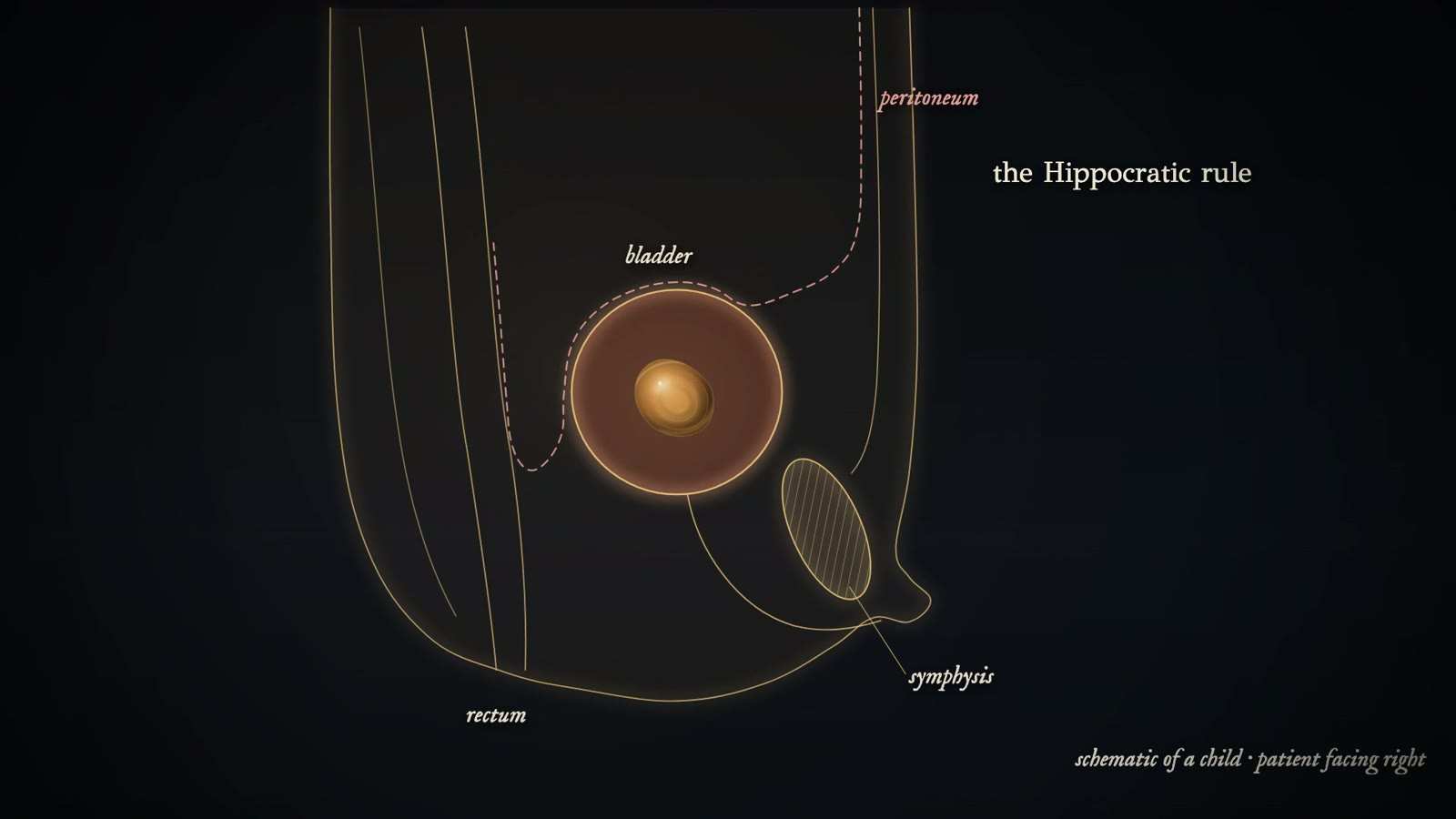
The bladder is opened through an incision above the pubic bone instead of through the perineum. Franco used it once, on a child whose large stone could not be drawn from below, against the Hippocratic teaching that wounds of the bladder were fatal, and did not recommend it. Rousset of Montpellier described it and practised it on the cadaver around 1581 to 1590. When it returned in early eighteenth-century London, Cheselden first distended the bladder with warm barley water run in through a catheter joined to an ox's ureter, so that it could be opened above the pubis; he later gave it up for the lateral operation.

Wooden music box with a gold cylinder and a label reading Frère Jacques, beneath a treble-clef staff with a row of notes.

Lateral perineal lithotomy

Period
Frère Jacques from 1697; Rau; Cheselden from 1725 to 1727
In the film
First appears 28:46 in the film, VI. The Stone-Cutters

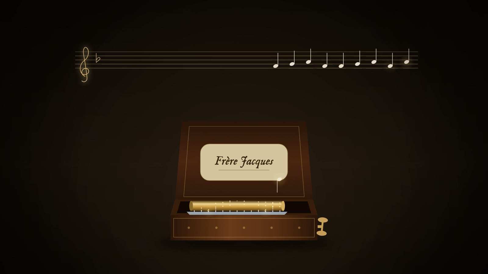
The incision is made in the perineum to the left of the midline raphe, and the bladder is entered through the side of its neck instead of along the urethra. Frère Jacques at first cut without a guide, and witnesses said there was no part of the region he had not cut. Rau and Cheselden used a grooved staff in the urethra, cutting onto it and following it into the bladder neck. Cheselden's instruments included the curved grooved staff, a short knife with a convex blade, a gorget that slid along the staff's groove, and stone forceps. The patient was bound in the lithotomy position and held by assistants. Cheselden's series gave about 9 per cent mortality.

Gold-line bladder with an amber stone held by three curved claws on a straight steel tube entering from the right, labelled the trilabe.

Lithotrity (crushing the stone through the urethra)

Period
From Civiale's first operation in January 1824 to Bigelow's litholapaxy of 1878
In the film
First appears 33:47 in the film, VII. Breaking the Stone

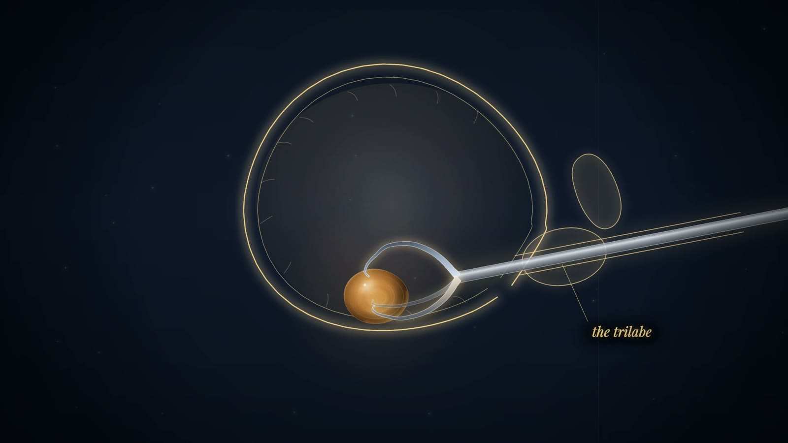
A steel instrument was passed along the urethra into the bladder, the stone was caught, and it was broken by drilling (Civiale's trilabe), by percussion (Heurteloup's percuteur) or, later, by blades closed with a screw. The fragments were left to pass with the urine. Following Civiale's teaching that the bladder tolerated instruments badly, each sitting lasted a few minutes, and a large stone could need many sittings over weeks: one patient in 1834 needed 37. After 1846 anaesthesia could be given; Thompson's operations on Napoleon III in 1873 were under chloroform.

Dim room with a surgeon bending over a patient beside a candle, under a quotation beginning My own conviction is that it is better to protract the operation.

Litholapaxy (rapid lithotrity with evacuation)

Period
Bigelow, 1878
In the film
First appears 37:14 in the film, VII. Breaking the Stone

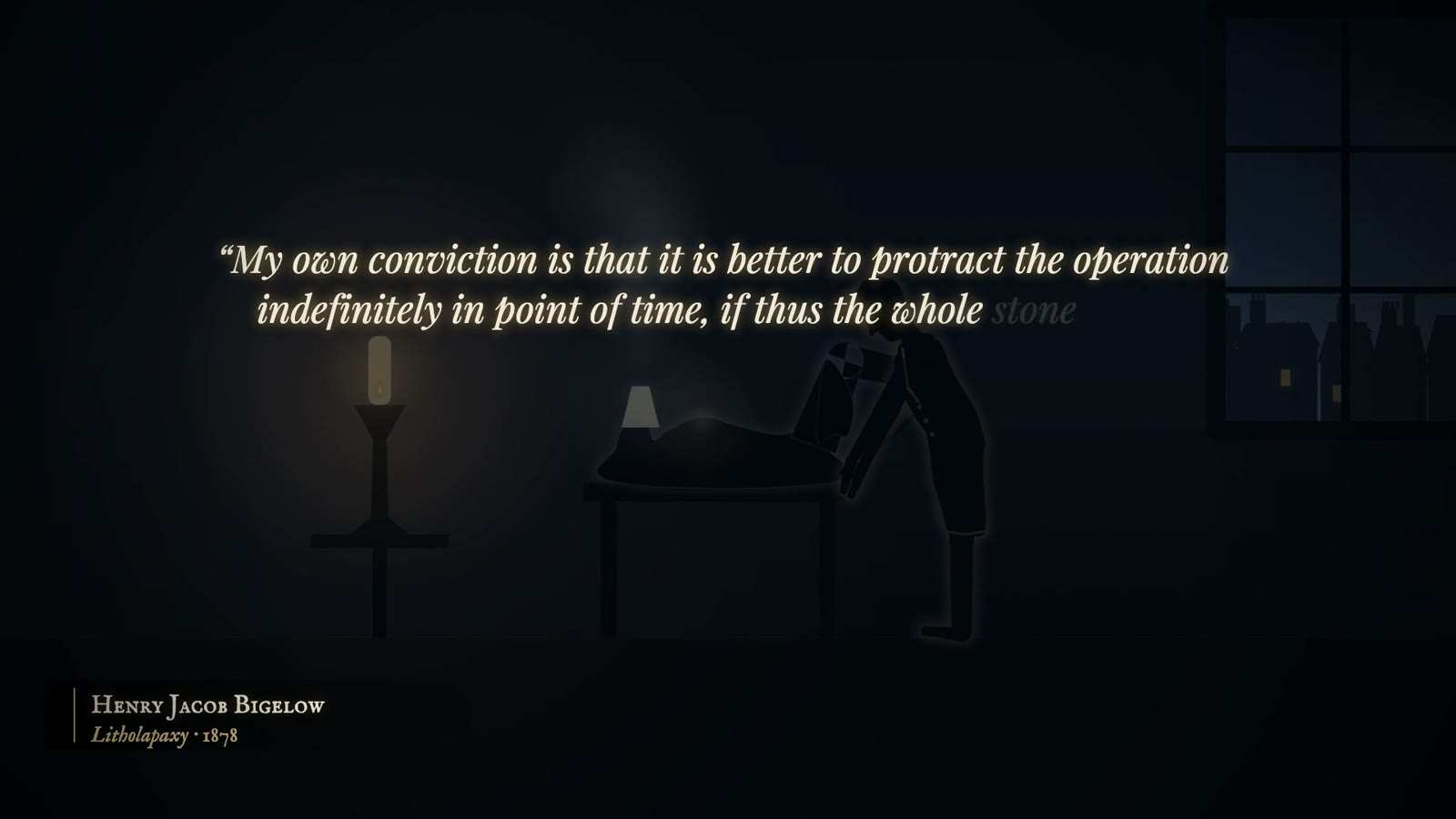
Under ether, the stone was crushed completely at a single sitting with a heavy lithotrite, and every fragment was then washed out through a wide evacuating catheter with Bigelow's rubber bulb and glass trap. Bigelow argued that the bladder tolerated a long operation far better than Civiale had taught, and that fragments left behind caused irritation and infection. His longest sitting lasted three and three-quarter hours; in one case he removed 706 grains of stone in a single sitting.

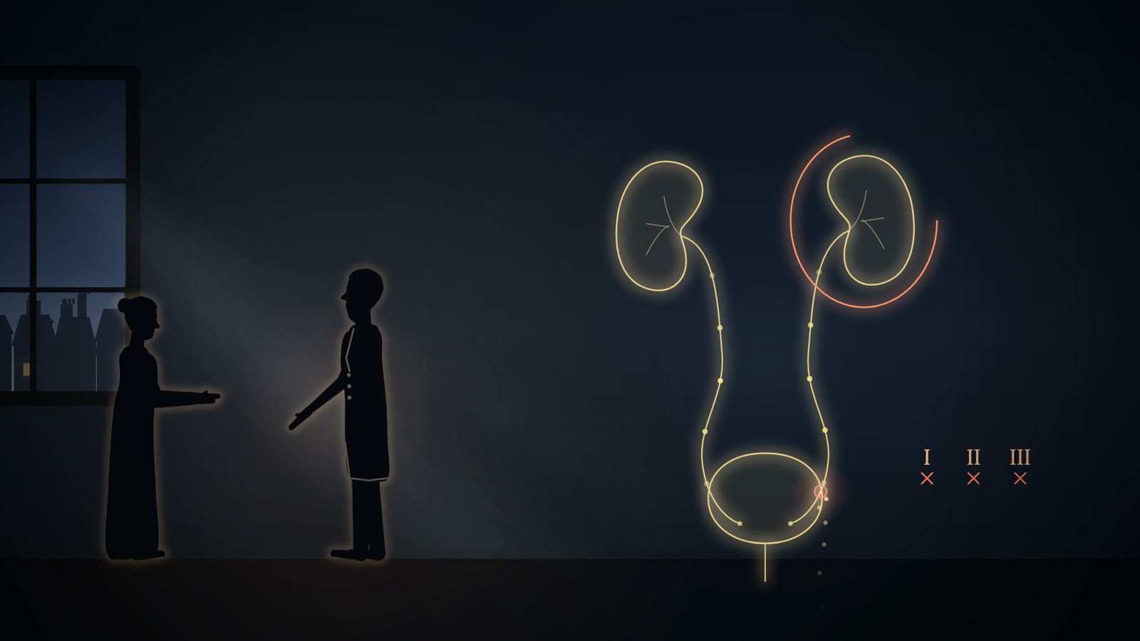
Gold-line kidneys, ureters and bladder with one kidney circled in red and tally marks I, II and III, beside two silhouetted figures by a window.

Planned nephrectomy (Simon, 1869)

Period
2 August 1869, Heidelberg
In the film
First appears 38:10 in the film, VII. Breaking the Stone

After practising on cadavers and removing a kidney from each of thirty dogs, Simon operated on Margaretha Kleb under chloroform through a lumbar incision. The kidney came out in forty minutes, with an estimated blood loss of 50 ml. Antisepsis was not yet routine: she developed a wound infection, pneumonia and erysipelas, but left her bed on day 28 and left hospital after two months. The aim was to stop a urinary fistula caused by an injured ureter, not to treat stone or tumour.

Sources
  1. BAUS Virtual Museum: Nephrectomy Museum or society
  2. Goddard and Birks, A short history of nephrectomy (Urology News) Secondary
  3. Wikipedia, Gustav Simon (surgeon) Secondary
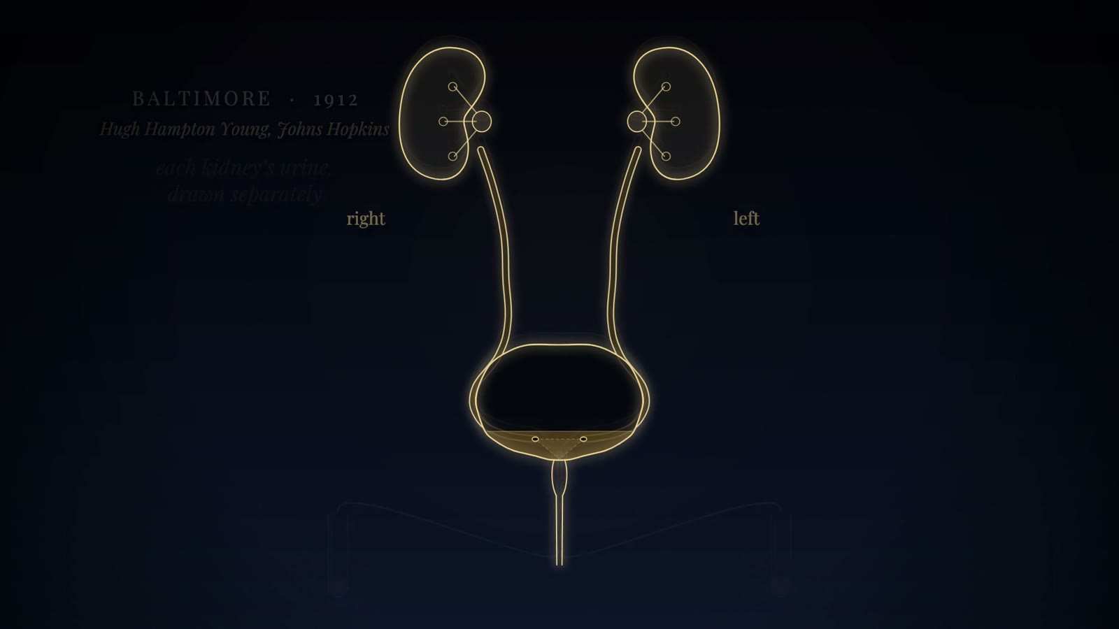
Gold-line kidneys, ureters and bladder with both ureteric openings marked, labelled right and left, with the caption Baltimore, 1912.

Cystoscopic ureteral catheterisation probable

Period
From the 1870s in women; steerable with Albarrán's lever from c. 1897
In the film
First appears 43:24 in the film, VIII. Light

A thin catheter was guided under vision into the opening of a ureter. Josef Grünfeld in Vienna, from 1873, is credited with the first such catheterisation under endoscopic control, in women, using external light reflected by a head mirror. David Newman of Glasgow saw and catheterised the ureters in 1883 with an electric bulb in the female bladder beside an open speculum. Karel Pawlík in Prague taught Howard Kelly in 1888 to catheterise the ureters through a speculum and to examine an air-filled bladder. Albarrán's lever then let the operator steer the catheter from within the cystoscope itself.

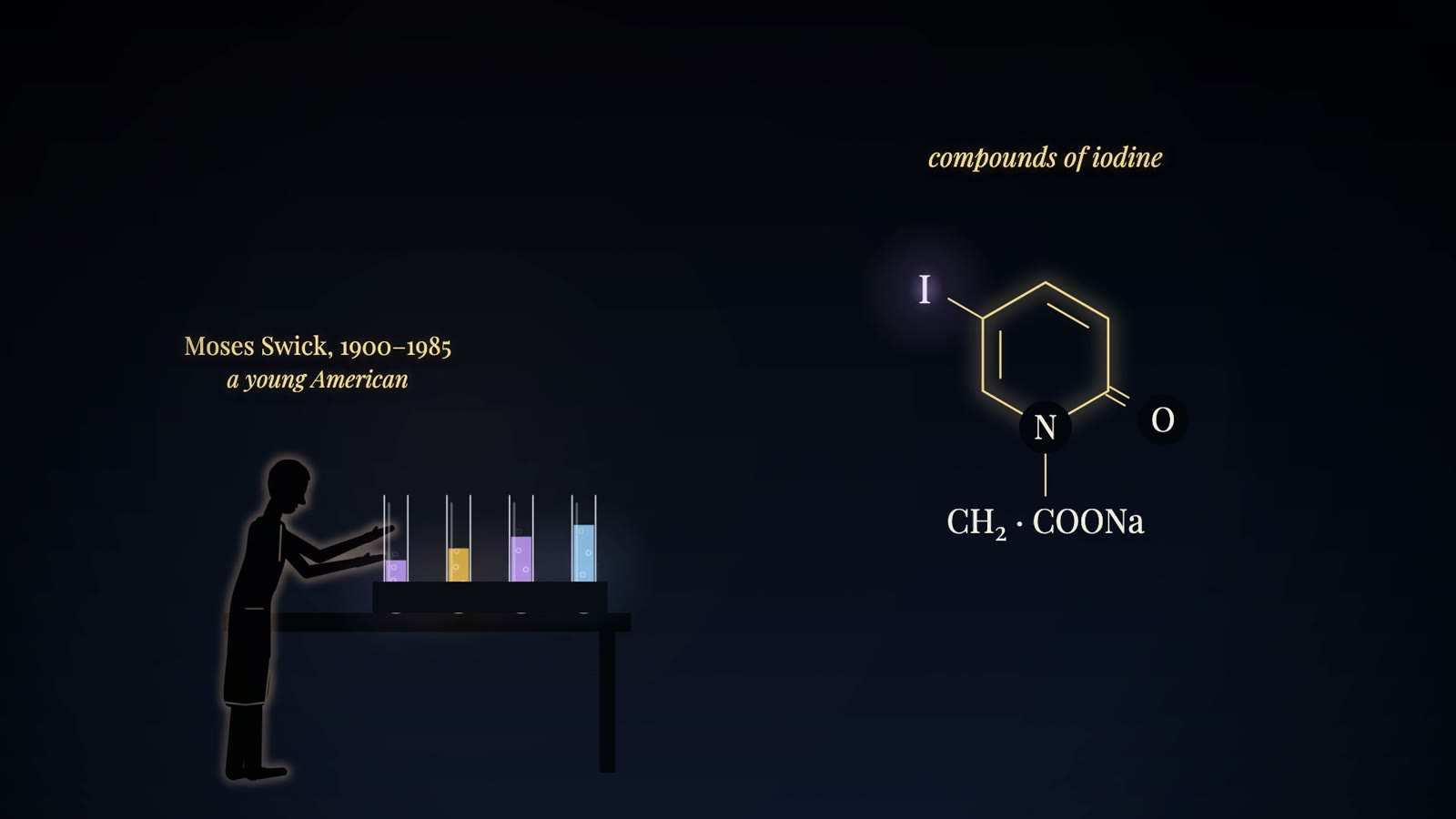
Silhouetted young chemist at a bench with four tubes of coloured liquid, beside a glowing structural formula with an iodine atom, captioned compounds of iodine.

Intravenous (excretory) urography with Uroselectan

Period
From 1929 until CT displaced it around the turn of the 21st century
In the film
First appears 45:23 in the film, VIII. Light

A contrast agent injected into a vein was carried to the kidneys and excreted in the urine, so a radiograph showed the kidneys, ureters and bladder without cystoscopy or catheterisation. The agent came from iodinated pyridine compounds that Arthur Binz and Curt Räth had made as anti-infectives. Moses Swick found that they were excreted rapidly by the kidney, and an iodinated N-acetylated derivative became Uroselectan: chemically, the sodium salt of 5-iodo-2-pyridone-N-acetic acid. The formula drawn in the film shows the N-acetate side chain as CH₂·COONa, confirmed at full resolution in the production review.

Silhouetted young chemist at a bench with four tubes of coloured liquid, beside a glowing structural formula with an iodine atom, captioned compounds of iodine.

Retrograde pyelography with Collargol

Period
Introduced 1906 at Heidelberg
In the film
First appears 45:23 in the film, VIII. Light

Through a cystoscope a ureteral catheter was passed up the ureter to the kidney, and the renal pelvis was filled with Collargol, a colloidal silver preparation, before a radiograph was taken. The plate showed the outline of the pelvis that the silver filled. Friedrich Voelcker and Alexander von Lichtenberg introduced the method. Every examination needed cystoscopy and ureteral catheterisation, and Collargol was an irritant; specific figures for its harms were not verified for the film. Intravenous urography removed the need for the cystoscope in 1929.

Sources
  1. Wikipedia (de): Friedrich Voelcker Secondary
  2. Wikipedia (de): Alexander von Lichtenberg Secondary
  3. Moll F, Friedrich Voelcker, Alexander v. Lichtenberg: Pyelographie, Aktuelle Urologie 1995;26:70-74, doi:10.1055/s-2008-1057777 Secondary
  4. Urology News: Early British pioneers of urological imaging Secondary
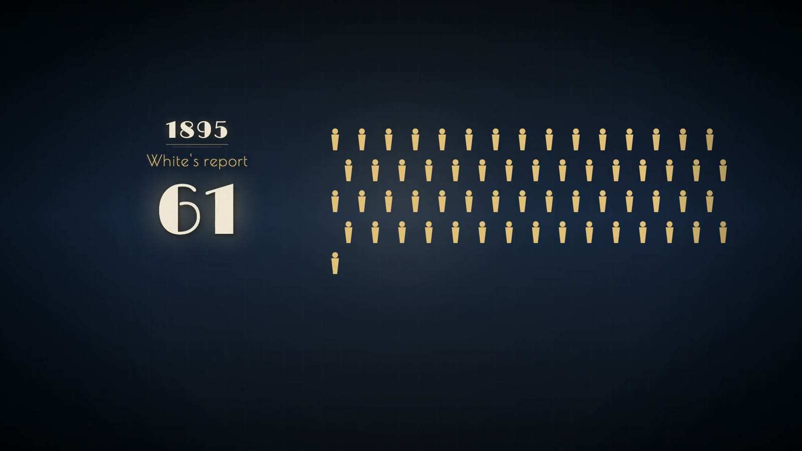
Date 1895 and a report caption above a counter reading 61, beside rows of small gold figures of men.

Castration for prostatic enlargement probable

Period
1893 to about 1900
In the film
First appears 49:24 in the film, IX. The Gland That Stands Before

Removal of both testes, offered to older men whose urine was obstructed by an enlarged prostate. The reasoning was an analogy: removing the ovaries shrinks fibroids of the womb, so removing the testes should shrink the prostate. In castrated dogs the gland shrank. In White's 1895 collection of 111 men the prostate shrank rapidly in 87% and symptoms improved in 83%, but 18% died. Vasectomy and ligation of the vas were tried as lesser versions. Enucleation of the gland replaced the operation within a few years.

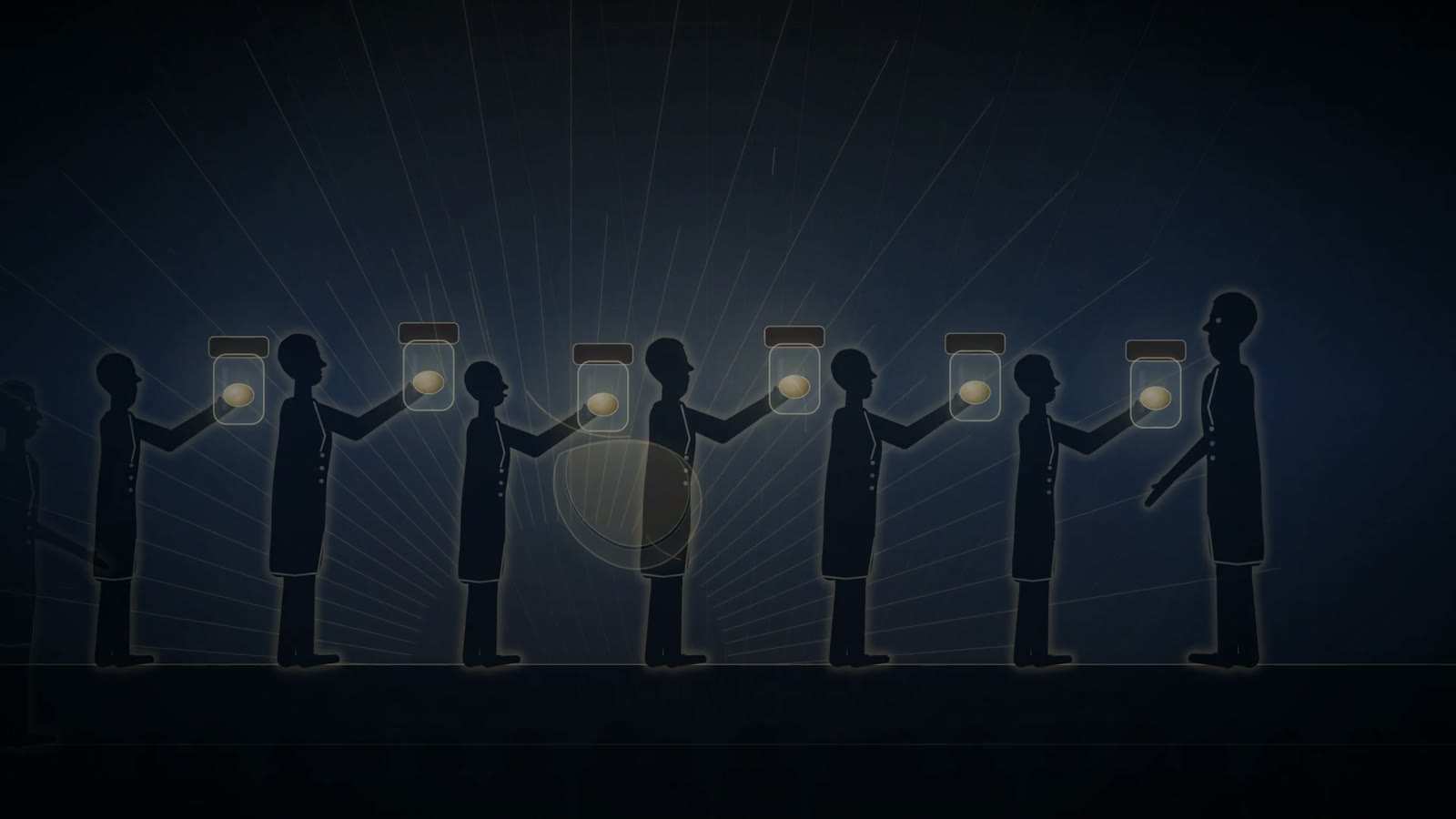
Row of silhouetted men in coats each holding up a glass specimen jar containing a pale oval specimen, with faint rays behind them.

Suprapubic (transvesical) prostatectomy

Period
1887 onward
In the film
First appears 50:12 in the film, IX. The Gland That Stands Before

The bladder is opened above the pubis and the enlarged gland is shelled out with a finger from inside the bladder. McGill removed prostatic tissue this way in Leeds in March 1887 and achieved complete enucleation that December; Belfield in Chicago did the same in the same period. Fuller of New York published a clear method of complete enucleation in 1895. Freyer, from December 1900, made it a standard operation, reporting more than a thousand cases, with mortality of about 3% in his last 200 by one account and 5% overall by another.

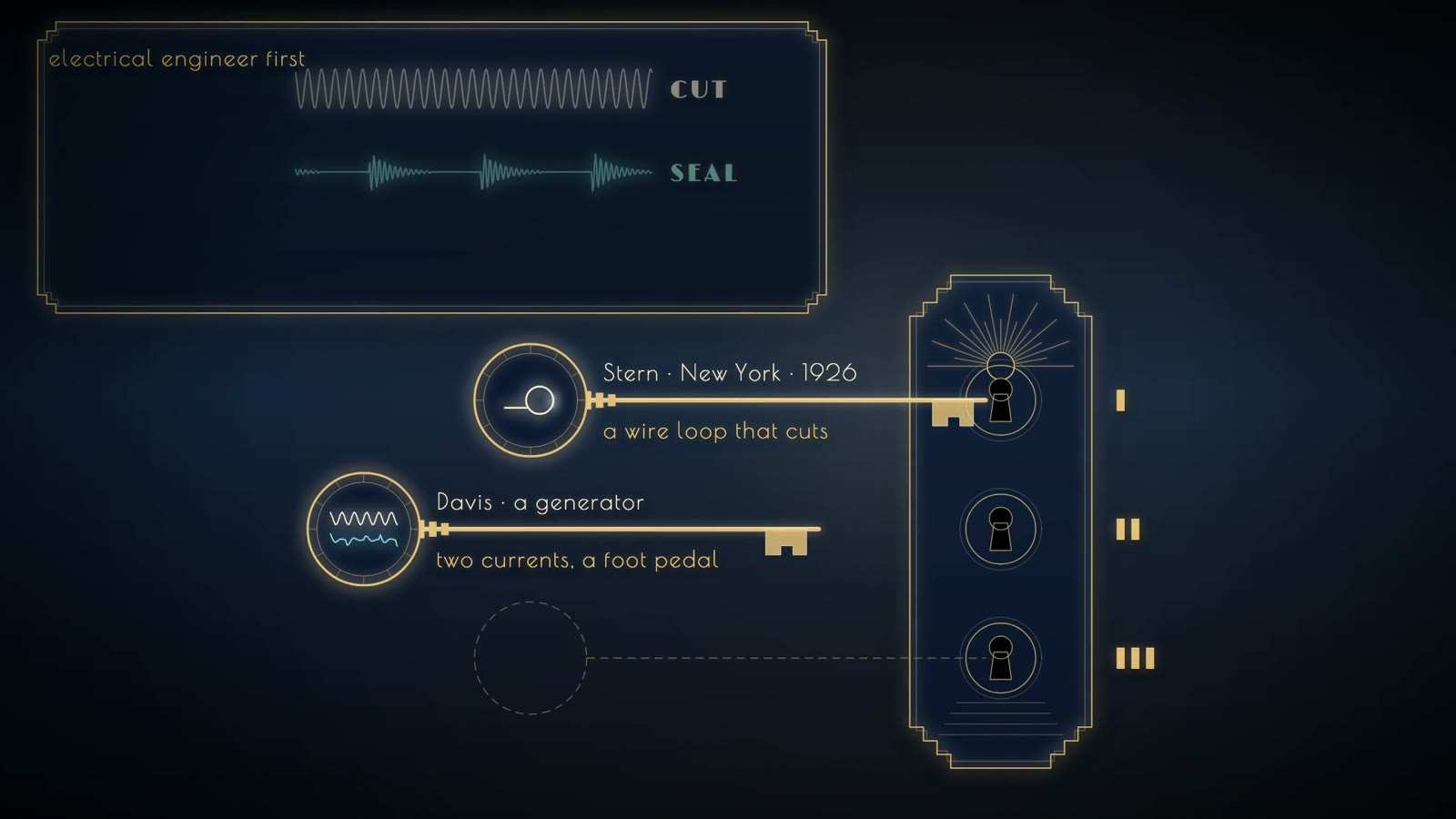
Lock with three keyholes; keys labelled Stern, New York, 1926, a wire loop that cuts, and Davis, a generator with two currents and a foot pedal; third empty.

Transurethral resection of the prostate (TURP)

Period
from 1931
In the film
First appears 51:07 in the film, IX. The Gland That Stands Before

Working through the urethra, with no incision in the skin, the surgeon views the obstructing prostate through the lens of the resectoscope. A wire loop carrying a high-frequency cutting current is drawn back through the tissue to remove it in strips, and a second, coagulating current, chosen with a foot switch, seals bleeding vessels. The method needed Stern's loop (1926), Davis's two-current generator and McCarthy's insulated, lensed sheath (1931). It avoided the abdominal or perineal wound and suited men too frail for open surgery; Davis reported 966 resections with 7 deaths.

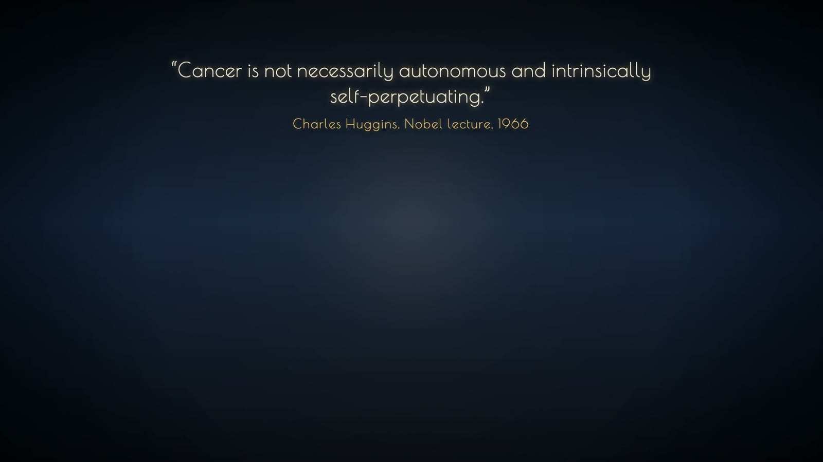
Quotation on a dark blue ground: Cancer is not necessarily autonomous and intrinsically self-perpetuating, attributed to a Nobel lecture, 1966.

Castration or oestrogen for advanced prostate cancer

Period
from 1941
In the film
First appears 53:08 in the film, IX. The Gland That Stands Before

Removal of the testes (orchiectomy), or oestrogen, withdraws the male hormone on which prostatic tissue depends. In 1941 Huggins and Hodges gave this treatment to men with metastatic prostate cancer and followed the serum acid phosphatase, which fell; injected androgen made it rise. Bone pain eased. Of the first 21 men treated by orchiectomy for far-advanced disease, 4 lived more than 12 years. Later, Schally's isolation of LHRH (structure confirmed 1971) led to agonist drugs that block the pituitary signal for sex steroids; the first trial in advanced prostate cancer, with Tolis in Montreal, was in 1982.

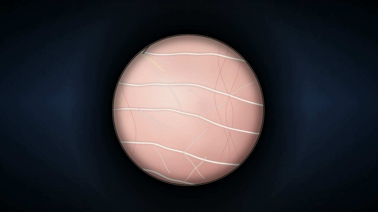
Round microscope field showing fine white nerve threads branching across a pale pink ground, with thin red vessels crossing on the right.

Nerve-sparing radical prostatectomy

Period
first performed 26 April 1982
In the film
First appears 55:40 in the film, IX. The Gland That Stands Before

Radical prostatectomy removes the whole prostate for cancer. Walsh first made it less bloody by identifying and tying the common venous trunk over the urethra. After he and Donker had traced the nerves of erection in 1981 and found them outside the prostate rather than within it, he designed the operation to leave them in place, separating them from the gland instead of removing them with it. The first patient, a 52-year-old professor of management, recovered potency quickly and remained free of cancer for decades.

Four silhouetted figures in two facing pairs, labelled urologist and surgeon; a small glowing kidney passes from one central figure to the other's abdomen.

Living-donor kidney transplantation between identical twins

Period
23 December 1954
In the film
First appears 58:33 in the film, X. Without the Knife

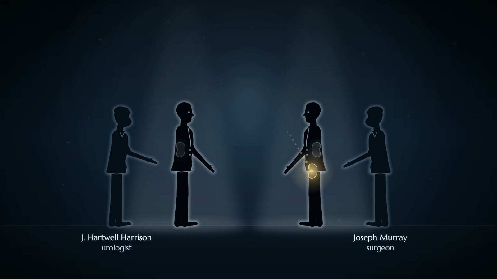
Before the operation, skin was grafted from each twin to the other; the grafts established that the brothers were genetically identical. The urologist Harrison removed a healthy kidney from the donor, Ronald Herrick, and Murray placed it in his brother Richard, who had kidney failure; Merrill's nephrology team cared for the recipient. The kidney worked at once, and Richard lived eight more years. The operation took about five and a half hours. It was the first time, Murray wrote, that a healthy person underwent major surgery not for his own benefit.

Water tank captioned Munich, with a gold curved reflector at its base and a kidney holding an amber stone above it, joined by a faint dashed ellipse.

Extracorporeal shock wave lithotripsy (ESWL)

Period
first patient 7 February 1980
In the film
First appears 1:00:03 in the film, X. Without the Knife

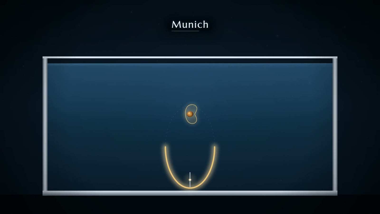
Shock waves made outside the body are focused on a kidney stone to break it without an incision. In the Dornier method an underwater spark at one focus of a half-ellipsoid produced the wave, which the reflector concentrated on the second focus; the stone was placed there under two-plane X-ray guidance while the patient lay in a water bath. Early treatment in Munich was limited to 206 strictly selected patients. The first lithotripsy centre opened in Munich on 20 May 1982, and the US FDA approved the device in December 1984.

Sources
  1. Chaussy, Eisenberger and Forssmann 2007, Extracorporeal shockwave lithotripsy (ESWL): a chronology (J Endourol 21:1249-1253) Primary
  2. Xu and Xu, history of shock wave lithotripsy (Atlantis Press) Secondary
  3. Storz Medical: 40 years of ESWL Secondary
  4. Malinaric et al., International Journal of Environmental Research and Public Health 2023; 20(5):4127 Secondary
Outline of two kidneys, ureters and bladder, an amber stone in one kidney and a thin scope entering the bladder from below, captioned a flexible ureteroscope.

Percutaneous nephrolithotomy (PCNL)

Period
1976 onward
In the film
First appears 1:00:55 in the film, X. Without the Knife

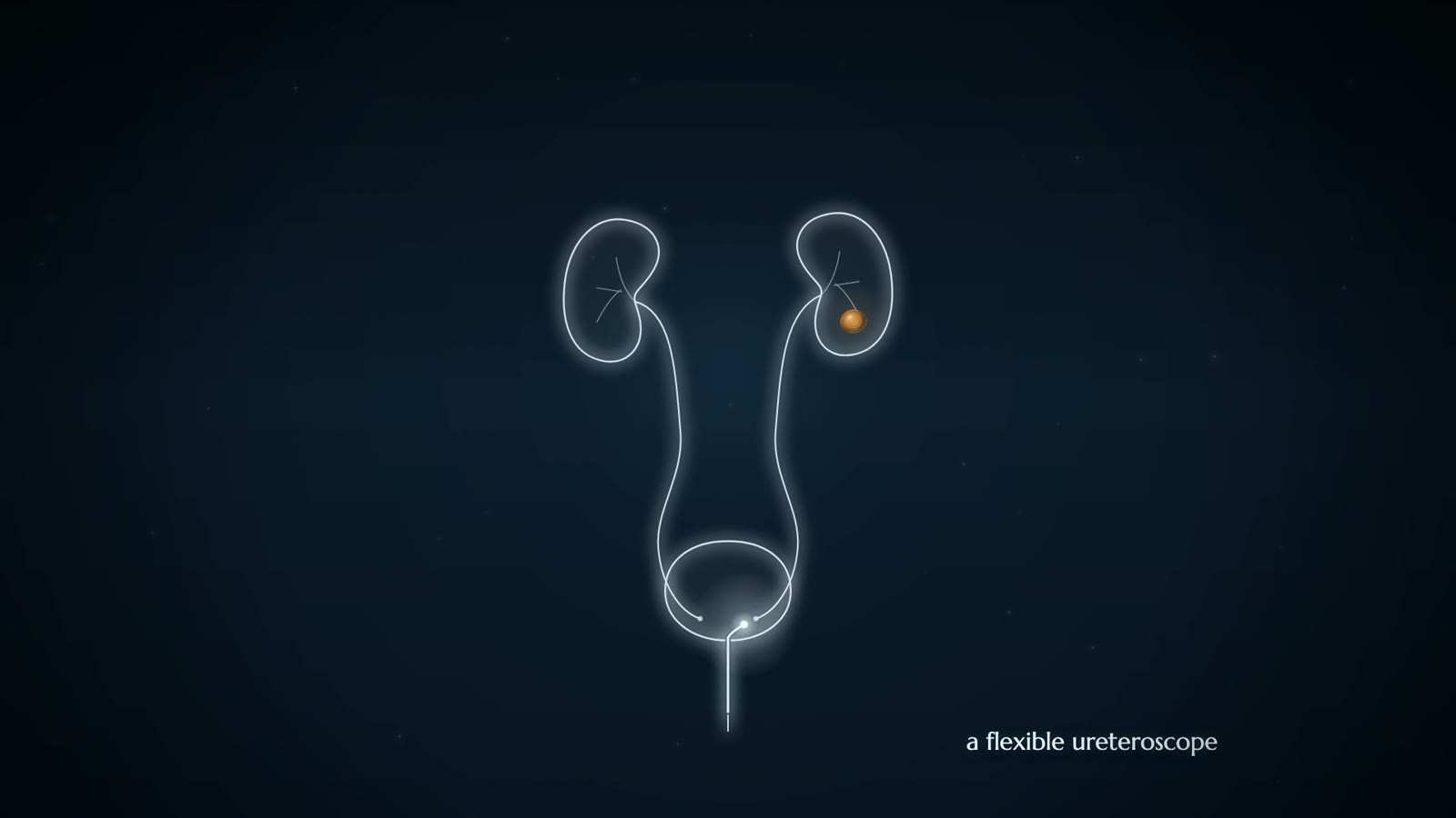
A track is made through the skin of the back into the collecting system of the kidney, and the stone is removed through it. Rupel and Brown had removed a stone under vision through a surgical nephrostomy track in 1941, and Goodwin described percutaneous nephrostomy in 1955. In 1976 Fernström and Johansson described extracting renal stones through a percutaneous nephrostomy under radiological control. Alken and colleagues and Wickham developed the technique, and Arthur Smith coined the word 'endourology' in 1978.

Sources
  1. BAUS Virtual Museum: Nephroscopy and PCNL Museum or society
  2. S. R. Patel, S. Y. Nakada, The modern history and evolution of percutaneous nephrolithotomy, Journal of Endourology 2014 Secondary
Hospital building in silhouette at night with lit windows and golden sparks gathering over its centre, captioned Henry Ford Hospital, Detroit.

Laparoscopic nephrectomy

Period
1990
In the film
First appears 1:01:15 in the film, X. Without the Knife

The kidney is freed inside the abdomen and removed through small punctures rather than an open incision. In the first case, at Washington University in St Louis in 1990, Clayman's team operated on an 85-year-old woman with a tumour in the right kidney. The freed kidney, 190 g, was placed in an entrapment sack inside her, broken up with an electrical morcellator and drawn out through an incision of 11 mm. The work was published in the Journal of Urology and the NEJM in 1991.

Sources
  1. R. V. Clayman et al., Journal of Urology 1991; 146:278-282, and New England Journal of Medicine 1991; 324:1370-1371 (Europe PMC abstract, PMID 1830346) Primary
  2. Wikipedia: Ralph Clayman Secondary
Hospital building in silhouette at night with lit windows and golden sparks gathering over its centre, captioned Henry Ford Hospital, Detroit.

Laparoscopic radical prostatectomy

Period
1991 to 1995 (first series)
In the film
First appears 1:01:15 in the film, X. Without the Knife

Removal of the whole prostate for cancer through laparoscopic ports instead of an open incision. Schuessler and colleagues, working with Clayman and Kavoussi, operated on nine men between September 1991 and May 1995. The mean operating time was 9.4 hours. In 1997 they concluded that the approach offered no advantage over open surgery in tumour removal, continence, potency, length of stay, convalescence or cosmetic result. Robotic assistance later revived minimally invasive removal of the prostate.

Sources
  1. W. W. Schuessler, P. G. Schulam, R. V. Clayman, L. R. Kavoussi, Urology 1997; 50:854-857 (Europe PMC abstract, PMID 9426713) Primary
  2. Wikipedia: Radical prostatectomy Secondary
Hospital building in silhouette at night with lit windows and golden sparks gathering over its centre, captioned Henry Ford Hospital, Detroit.

Robot-assisted radical prostatectomy

Period
from 2000
In the film
First appears 1:01:15 in the film, X. Without the Knife

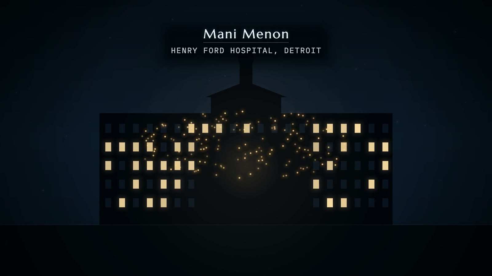
Radical prostatectomy performed laparoscopically with a robotic system; in the film's description the surgeon works at a console while mechanical arms hold the instruments. Binder and Kramer performed the first in Frankfurt in 2000 and reported it in BJU International in 2001. Mani Menon's programme at the Henry Ford Hospital in Detroit, described as the first cancer-oriented robotics programme, developed it further. Its adoption outran high-quality evidence of advantage over open surgery (Lowrance et al. 2010). A single-port system was cleared for urology in 2018.

Sources
  1. J. Binder, W. Kramer, Robotically-assisted laparoscopic radical prostatectomy, BJU International 2001; 87:408-410 Primary
  2. Wikipedia: Robot-assisted surgery Secondary
  3. Wikipedia article on Mani Menon, as quoted in research/05 §10.1 (URL not recorded in the kit) Secondary
  4. W. T. Lowrance et al. 2010, abstract read on Europe PMC (PMID 21103791) Primary
  5. FDA 510(k) K173906: da Vinci SP (2018) Primary